Skip to content
DSD Research Library Public Research Platform
English
  • 简体中文
  • English
  • Français
  • Русский
  • Español
  • Português
Search Cases Research Atlas Papers Learn Methods

Case image gallery

Browse cases and figures from the original collection. Search by condition, gene, karyotype or phenotype and follow the source publication.

280 Image cases571 Images

Case recordsCase image gallery 280
All

Current selection: 1 Image cases · 1 Images

Select an image to enlarge it. Original images retain the complete source file.

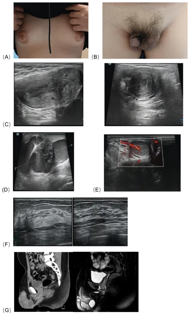
FIGURE 1 (A) Bilateral breast development, (B) The patient ’ s external genitalia, (C) Ultrasonography revealed an abnormal solid mass in the left inguinal region and scrotum, consistent with a uterus, measuring approximately 5.7×3.0×2.4 cm, with endometrial echo (thickness ~0.7 cm) and uterine cavity separation (~0.4 cm). (D) Surrounding the mass were tortuous, dilated tubular anechoic areas, suggestive of fallopian tubes, measuring approximately 7.1×4.2×6.1 cm. (E) The right scrotum contained testicular parenchyma, measuring 2.0×1.4×0.8 cm. (F) Breast ultrasound showed glandular tissue in both breasts, measuring 1.2 cm in anteroposterior diameter, with no detectable masses. (G) CT and MRI revealed a tubular soft tissue structure with a lumen, measuring approximately 4.8×1.5×1.1 cm, was detected between the bladder and rectum. 1 Images

DSD-C034

46,XX DSD · 46,XX · DSD-C034

46,XX · HPO:0000047, HPO:0000771, Hypospadias (HP:0000047), Gynecomastia (HP:0000771)

SRY-negative 46,XX testicular disorder of sex development with ectopic Müllerian structures; right gonadal biopsy confirmed testicular tissue.

CaseSource publicationAtlas

Swipe left or right to browse this case’s images

Open original image
CaseSource publicationAtlasView original caption in Chinese
DSD Research Library Data version v1.4.0