DSD-C016
46,XX DSD · 46,XX · DSD-C016
Not reported · Therefore, the leading hypothesis at diagnosis was CYP19 deficiency, based on severe maternal virilization during pregnancy. However, CYP19 gene analysis did not confirm this diagnosis
Images · 5
Case fields and evidence
- Category
-
46,XX DSD
- Age
-
Not reported
- Sex
-
Assigned male because of the virilized external genitalia appearance.
- Karyotype
-
46,XX
- Phenotypes
-
Hypogonadism (HP:0000135)
- Diagnosis
-
Not reported · Therefore, the leading hypothesis at diagnosis was CYP19 deficiency, based on severe maternal virilization during pregnancy. However, CYP19 gene analysis did not confirm this diagnosis
- Follow-up
-
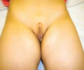
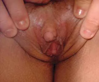
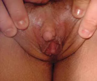
Mother (A) and patient (B) external genitalia 8 months after delivery. Laboratory follow-up from 8 to 105 months is detailed in the original.
- General phenotype
-
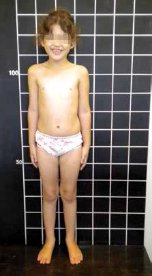
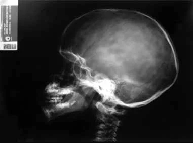
General examination showed virilized external genitalia, Prader stage V, penis 2.3 cm, non-palpable gonads; glanular urethral opening and complete fusion (POR deficiency and Antley-Bixler syndrome).
- Imaging
-
Pelvic ultrasound showed a 1.1 cm³ uterus and gonads of typical ovarian morphology, with an anechoic homogeneous shadow in the left adnexal region suggesting a simple ovarian cyst.
- Laboratory findings
-
Not reported
- Management
-
Apart from around two additional surgeries, hydrocortisone replacement was never required: at age 5, vaginal dilation for vaginal stenosis and endometritis; at age 6, clitoroplasty.
- Pathology
-
Gonadal biopsy confirmed the presence of two normal ovaries.
- Evidence level
-
See source
- Clinical history
-
See source
- Perinatal history
-
See source
- Family history
-
See source
- Growth and development
-
See source
- External genital examination
-
See source
- Genetics
-
See source
Cite this resource
DSD Research Library | 46,XX DSD · 46,XX · DSD-C016 | v1.4.0 | 2026-11-17 | https://dsdatlas.com/en/cases/DSD-C016?version=v1.4.0