DSD-C034
46,XX DSD · 46,XX · DSD-C034
SRY-negative 46,XX testicular disorder of sex development with ectopic Müllerian structures; right gonadal biopsy confirmed testicular tissue.
Images · 1
Case fields and evidence
- Category
-
46,XX DSD
- Age
-
18-year-old
- Sex
-
The patient was reared as a male from birth.
- Karyotype
-
46,XX
- Phenotypes
-
Hypospadias (HP:0000047); Gynecomastia (HP:0000771)
- Diagnosis
-
SRY-negative 46,XX testicular disorder of sex development with ectopic Müllerian structures; right gonadal biopsy confirmed testicular tissue.
- Follow-up
-
Postoperatively, the patient recovered well.
- General phenotype
-
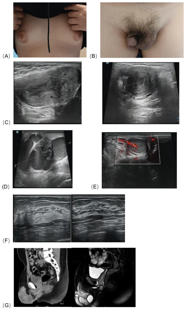
On physical examination, the patient’s vital signs were as follows: temperature 36.5 °C, blood pressure 112/65 mmHg, height 158 cm, weight 66.4 kg, and BMI 26.6 kg/m². Bilateral breast development was observed (Figure 1A), consistent with Tanner stage III.
- Imaging
-
Ultrasonographicexaminationofthescrotumandtestesrevealedan abnormal solid mass in the left hemiscrotum, which was suggestive of a uterus with possible intrauterine fluid accumulation, measuring approximately 5.7×3.0×2.4 cm (Figure 1C). Ultrasonography of the adrenal glands, liver, gallbladder, pancreas, spleen, and kidneys showed no abnormalities. Subsequent CT and MRI examinations revealed no gonadal tissue in the pelvic or abdominal cavity;
- Laboratory findings
-
Not reported
- Management
-
10.3389/fendo.2026.1829751 Frontiers in Endocrinology frontiersin.org 02 psychological evaluation, and approval by the ethics committee, we decided to proceed with the complete excision of the left scrotal mass while preserving the right testis and penis.
- Pathology
-
Diagnostic laparoscopy and management were performed under general anesthesia, including laparoscopic exploration and bilateral gonadal biopsy. Histological examination of the gonadal tissue samples from the right hemiscrotum, after formalin fixation, paraffin embedding, and hematoxylin-eosin staining, showed multiple seminiferous tubules interspersed within fibrous tissue, with thickened and fibrotic walls.
- Evidence level
-
See source
- Clinical history
-
See source
- Perinatal history
-
See source
- Family history
-
See source
- Growth and development
-
See source
- External genital examination
-
See source
- Puberty / Tanner
-
See source
- Genetics
-
See source
Cite this resource
DSD Research Library | 46,XX DSD · 46,XX · DSD-C034 | v1.4.0 | 2026-11-17 | https://dsdatlas.com/en/cases/DSD-C034?version=v1.4.0