DSD-C036
Other / unclassified DSD · 46,XY · DSD-C036
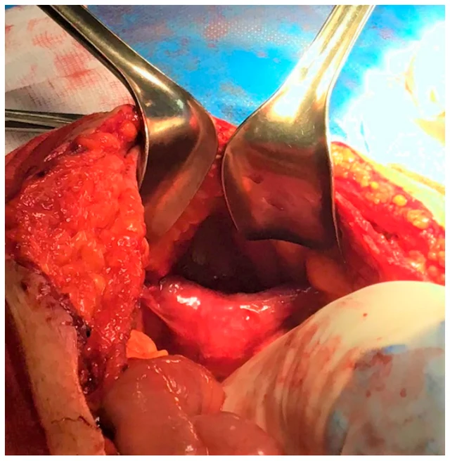
Based on the intraoperative findings, history of bilateral cryptorchidism, MRI imaging and karyotype, diagnosis of PMDS was made. It is very likely that this figure underestimates the true incidence of the syndrome as the diagnosis of PMDS largely depends on the intraoperative detection of Müllerian derivatives.
Images · 1
Case fields and evidence
- Category
-
Other / unclassified DSD
- Age
-
49-year-old
- Sex
-
Not reported
- Karyotype
-
46,XY
- Phenotypes
-
Hypospadias (HP:0000047); Cryptorchidism (HP:0000028)
- Diagnosis
-
Based on the intraoperative findings, history of bilateral cryptorchidism, MRI imaging and karyotype, diagnosis of PMDS was made. It is very likely that this figure underestimates the true incidence of the syndrome as the diagnosis of PMDS largely depends on the intraoperative detection of Müllerian derivatives.
- Follow-up
-
The subsequent imaging follow-up with testicular and pelvic ultrasound were negative for malignant testicular and Müllerian derivative degeneration.
- General phenotype
-
The MRI showed the presence of an 8.5 × 2 cm cylindrical, solid lesion, located above the bladder, similar to a normoflexed, rudimentary, dysmorphic uterus, probably originating from the prostatic urethra (Figure 2). Testicular echo-color-doppler was performed, confirming the mild bilateral testicular atrophy detected on physical examination, but no lesions suggestive of malignancy.
- Imaging
-
Preoperative imaging consisted of a CT scan limited to the upper abdominal wall, showing 9 × 11 cm diastasis of the anterior rectus abdominal muscles. The surgical findings were discussed with the patient, and he consented to a further diagnostic examination with a contrast enhanced MRI of the lower abdomen. The MRI showed the presence of an 8.5 × 2 cm cylindrical, solid lesion, located above the bladder, similar to a normoflexed, rudimentary, dysmorphic uterus, probably originating from the prostatic urethra (Figure 2).
- Laboratory findings
-
Not reported
- Management
-
Not reported
- Pathology
-
The patient must be reassured about his sexual identity and it must be explained that the pathology does not affect normal sexual function, although it is often associated with infertility.
- Evidence level
-
See source
- Clinical history
-
See source
- External genital examination
-
See source
Cite this resource
DSD Research Library | Other / unclassified DSD · 46,XY · DSD-C036 | v1.4.0 | 2026-11-17 | https://dsdatlas.com/en/cases/DSD-C036?version=v1.4.0