Skip to content
DSD Research Library Public Research Platform
English
  • 简体中文
  • English
  • Français
  • Русский
  • Español
  • Português
Search Cases Research Atlas Papers Learn Methods

Case image gallery

Browse cases and figures from the original collection. Search by condition, gene, karyotype or phenotype and follow the source publication.

280 Image cases571 Images

Case recordsCase image gallery 280
All

Current selection: 1 Image cases · 3 Images

Select an image to enlarge it. Original images retain the complete source file.

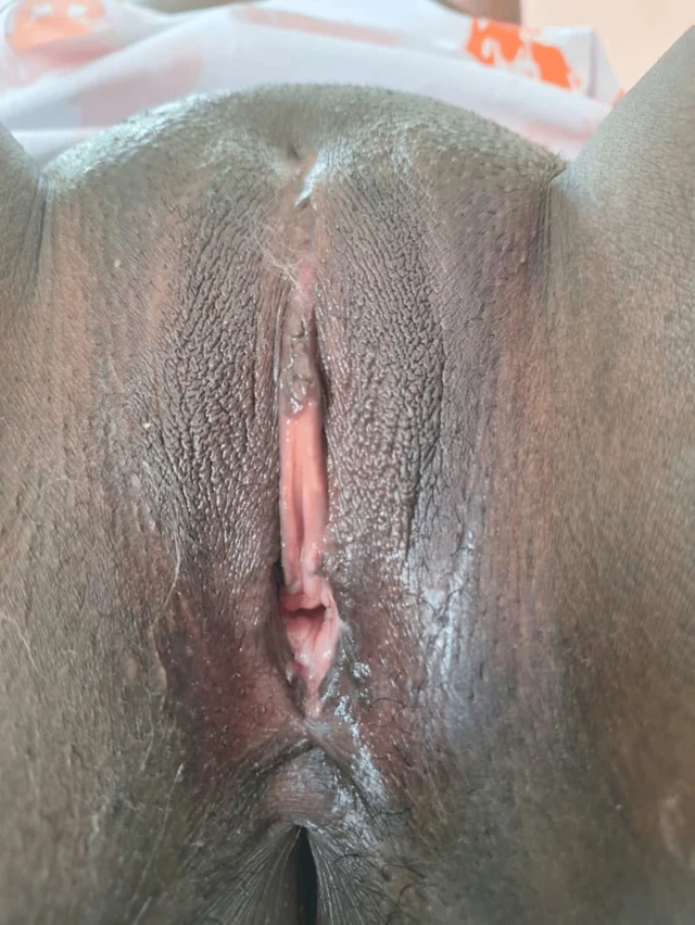
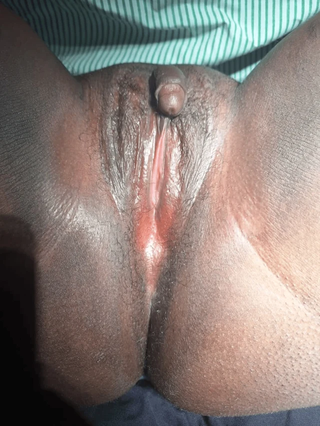
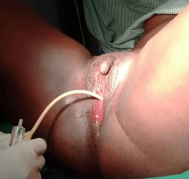
FIGURE 2: Case one catheterized At the six-week follow-up, the patient reported marked improvement in psychological well-being and satisfaction with the surgical outcome. Examination demonstrated a well-healed perineum and a functional vaginal canal measuring approximately 7 cm in length and 3 cm in width, as shown in Figure 3. She was also reviewed at three months and six months postoperatively, and she was happily sexually active without a complaint. Physical examination showed a long wide vaginal opening of about 7 cm long by 3 cm wide. 2026 Ainomugisha et al. Cureus 18(6): e111019. DOI 10.7759/cureus.111019 4 of 11 3 Images
FIGURE 3: Case one six weeks post-surgery Case two A 25-year-old individual raised and socially recognized as male presented with progressive abdominal distension and lower abdominal pain of one year duration. During clinical evaluation, the patient disclosed the onset of virilization during adolescence, including the development of facial hair and a deepened voice, but also reported regular menstruation beginning at 15 years of age. The patient had been assigned male gender at birth because of the presence of a phallus, which had enlarged progressively with age. However, urine did not pass through the phallus. The patient identified socially as male and expressed a desire for relief from the abdominal swelling and cessation of menstruation. Both parents were deceased, and the patient was accompanied by a community health worker in whom he had previously confided. He had no formal education and survived on casual labor. On examination, the patient appeared chronically ill and wasted but demonstrated masculine body habitus, facial hair, a hoarse voice, smooth skin, and well-developed breasts. Vital signs were stable. Abdominal examination revealed gross distension with marked suprapubic tenderness. A large solid pelvic mass measuring approximately 18 × 10 cm was palpable, in addition to another irregular mass within the left lumbar region. Bilateral inguinal nodular swellings were also noted. Perineal examination demonstrated a phallus measuring approximately 6 × 3 cm without a urethral opening. A closed introitus with a small distal dimple was observed, through which urine was passed with straining in the lithotomy position. The stage, according to the Prader score, was 2. The anal opening was normally positioned. A provisional diagnosis of 2026 Ainomugisha et al. FIGURE 1: Case one before surgery A provisional diagnosis of ambiguous genitalia with suspected 46, XX DSD was made. Pelvic ultrasonography demonstrated a normal uterus, cervix, and right ovary. A cystic lesion measuring 33 × 4 × 20 mm with multiple internal echoes and a thick wall was identified within the left ovary. No abnormalities were detected in the urinary tract or upper abdominal organs. Due to financial limitations, karyotyping and hormonal assays could not be performed. The patient expressed a strong desire for surgical correction of the vaginal obstruction and removal of the enlarged clitoris after extensive counseling regarding anatomy, sexual function, and potential risks of surgery. Surgical Procedure Under spinal anesthesia and lithotomy positioning, examination under anesthesia confirmed a normal urethral opening located superior to the vaginal dimple. Catheterization, as shown in Figure 2, verified that the enlarged clitoris was not associated with the urinary tract. A diamond-shaped incision was created at the vaginal opening to enlarge the introitus. A vaginal canal approximately 6 cm long and 2 cm wide was identified, and the cervix was visualized. Vaginal mucosa was mobilized and anchored to the labial and perineal skin edges. A 20-mL syringe barrel was inserted and secured as a temporary vaginal stent. Clitoral reduction was subsequently performed by excision at the base near the mons pubis, followed by reconstruction and layered closure with meticulous hemostasis. Postoperatively, the patient remained hospitalized for 10 days and received antibiotics, analgesics, and perineal hygiene care. The urinary catheter 2026 Ainomugisha et al. Cureus 18(6): e111019. DOI 10.7759/cureus.111019 3 of 11

DSD-C028

46,XX DSD · 46,XX · DSD-C028

46,XX

作者报告为生殖器歧义,疑46,XX DSD。

CaseSource publicationAtlas
Swipe left or right to browse this case’s images

Open original image
CaseSource publicationAtlasView original caption in Chinese
DSD Research Library Data version v1.4.0