46,XX DSD · 46,XX · DSD-C028
IMGc455277ffcd2d0 · 2892 × 3849
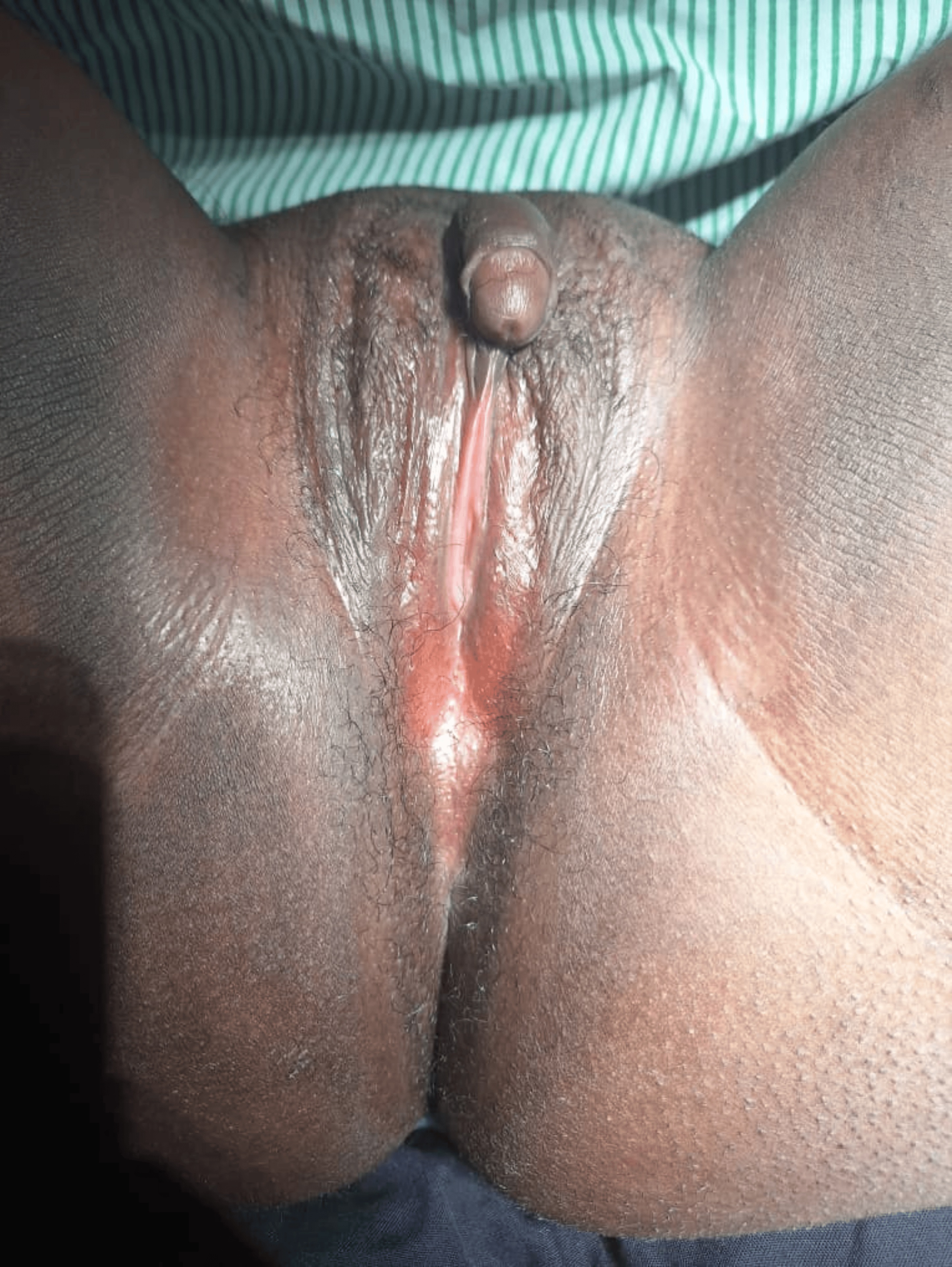
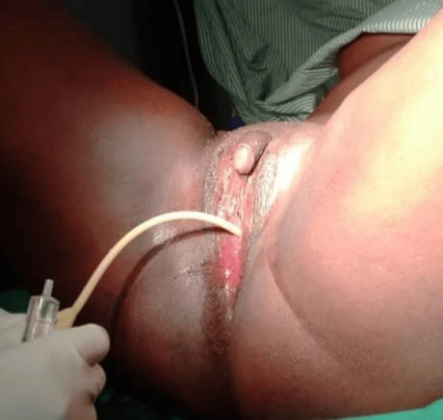
FIGURE 1: Case one before surgery A provisional diagnosis of ambiguous genitalia with suspected 46, XX DSD was made. Pelvic ultrasonography demonstrated a normal uterus, cervix, and right ovary. A cystic lesion measuring 33 × 4 × 20 mm with multiple internal echoes and a thick wall was identified within the left ovary. No abnormalities were detected in the urinary tract or upper abdominal organs. Due to financial limitations, karyotyping and hormonal assays could not be performed. The patient expressed a strong desire for surgical correction of the vaginal obstruction and removal of the enlarged clitoris after extensive counseling regarding anatomy, sexual function, and potential risks of surgery. Surgical Procedure Under spinal anesthesia and lithotomy positioning, examination under anesthesia confirmed a normal urethral opening located superior to the vaginal dimple. Catheterization, as shown in Figure 2, verified that the enlarged clitoris was not associated with the urinary tract. A diamond-shaped incision was created at the vaginal opening to enlarge the introitus. A vaginal canal approximately 6 cm long and 2 cm wide was identified, and the cervix was visualized. Vaginal mucosa was mobilized and anchored to the labial and perineal skin edges. A 20-mL syringe barrel was inserted and secured as a temporary vaginal stent. Clitoral reduction was subsequently performed by excision at the base near the mons pubis, followed by reconstruction and layered closure with meticulous hemostasis. Postoperatively, the patient remained hospitalized for 10 days and received antibiotics, analgesics, and perineal hygiene care. The urinary catheter 2026 Ainomugisha et al. Cureus 18(6): e111019. DOI 10.7759/cureus.111019 3 of 11
Images · 3