Aller au contenu
DSD Research Library Plateforme publique de recherche
Français
  • 简体中文
  • English
  • Français
  • Русский
  • Español
  • Português
Recherche Cas cliniques Recherche Atlas Publications Formation Méthodes

Retour à la galerie de cas

Dysgénésie gonadique · 45,X/46,XY · DSD-C042

IMG40da2b71a8f1ad · 1123 × 517
CasPublication sourceAtlasOuvrir l’image originale
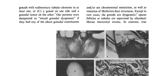
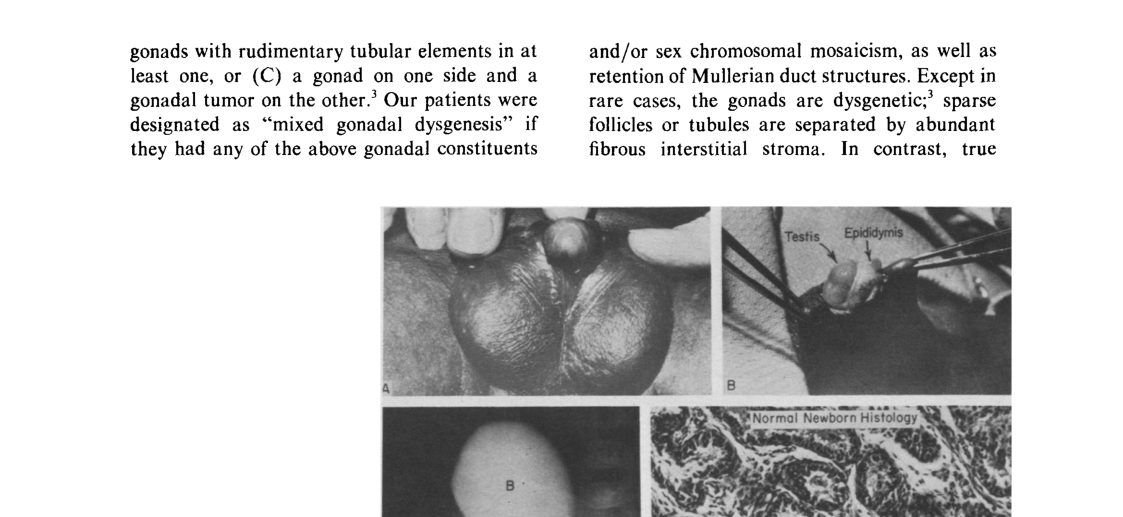
Fig. 5. Patient 13. This 45,X/47,XYY mosaic had deficient but symmetric (A) external genitalia and bilateral testes (B) in the scrotal folds. Vagina (C) alone remained of the Mullerian Duct structures. Testicular histology (D) was that of a normal newborn, despite the fact that the testosterone response to HCG was low.
Légende

Fig. 5. Patient 13. This 45,X/47,XYY mosaic had deficient but symmetric (A) external genitalia and bilateral testes (B) in the scrotal folds. Vagina (C) alone remained of the Mullerian Duct structures. Testicular histology (D) was that of a normal newborn, despite the fact that the testosterone response to HCG was low.

Images · 5
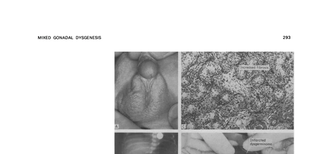
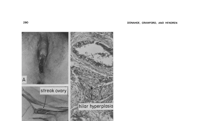
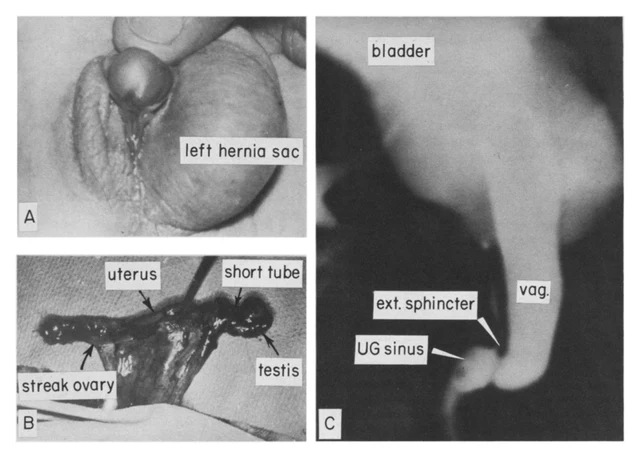
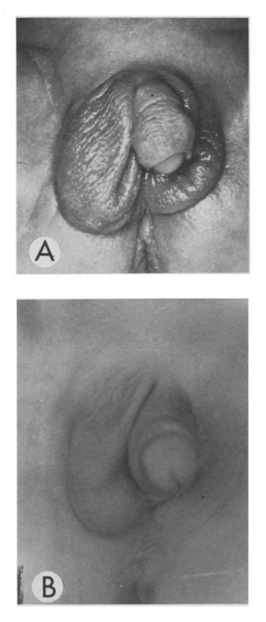
Fig. 6. Patient 11. This newborn mosaic 45,X/46,XY has (A} deficient external genitalia. A mass in the left abdomen (B) displaced bowel to the right. (C) The testicular histology on the right showed slight increase in interstitial fibrosis. (D) The gonad on the left twisted on a narrow stalk and infarcted. Fig. 2. Patient 5. (A) Modest clitoral hypertrophy occurred at age 13. (B) The streak ovary showed histologic evidence of (C) hilar hyperplasia. Fig. 5. Patient 13. This 45,X/47,XYY mosaic had deficient but symmetric (A) external genitalia and bilateral testes (B) in the scrotal folds. Vagina (C) alone remained of the Mullerian Duct structures. Testicular histology (D) was that of a normal newborn, despite the fact that the testosterone response to HCG was low. Fig. 1. Patient 1. (A) External genitalia with hernia uteri inguinale on the left and perineoscrotal hypospadias. (B) Asymmetric gonads--dysgenetic testis and streak ovary, The truncated follopian tube on the side of the testis demonstrates the local but diminished influence of Mullerian Inhibiting Substance. (C) Sinogram fills a vagina that enters the urogenital sinus just distal to the external urethral sphincter, Fig. 3. Patient 10. External genitalia showed very little development from birth (AI to age 6 (B).

Balayez à gauche ou à droite pour voir les images de ce cas

Ouvrir l’image originale
CasPublication sourceAtlasVoir la légende originale en chinois
DSD Research Library Version des données v1.4.0