Ir para o conteúdo
DSD Research Library Plataforma pública de pesquisa
Português
  • 简体中文
  • English
  • Français
  • Русский
  • Español
  • Português
Pesquisa Casos Investigação Atlas Publicações Educação Métodos

Voltar à galeria de casos

DSD 46,XX (diferenças do desenvolvimento sexual) · 46,XX · DSD-C034

IMG022a16065766de · 641 × 1067
CasoPublicação originalAtlasAbrir imagem original
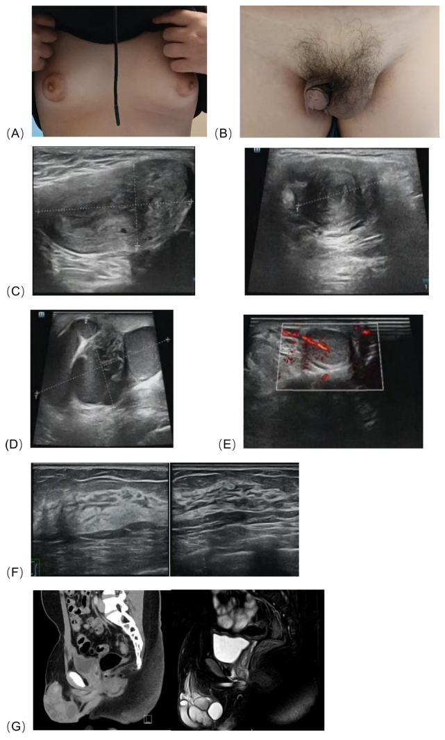
FIGURE 1 (A) Bilateral breast development, (B) The patient ’ s external genitalia, (C) Ultrasonography revealed an abnormal solid mass in the left inguinal region and scrotum, consistent with a uterus, measuring approximately 5.7×3.0×2.4 cm, with endometrial echo (thickness ~0.7 cm) and uterine cavity separation (~0.4 cm). (D) Surrounding the mass were tortuous, dilated tubular anechoic areas, suggestive of fallopian tubes, measuring approximately 7.1×4.2×6.1 cm. (E) The right scrotum contained testicular parenchyma, measuring 2.0×1.4×0.8 cm. (F) Breast ultrasound showed glandular tissue in both breasts, measuring 1.2 cm in anteroposterior diameter, with no detectable masses. (G) CT and MRI revealed a tubular soft tissue structure with a lumen, measuring approximately 4.8×1.5×1.1 cm, was detected between the bladder and rectum.
Legenda da figura

FIGURE 1 (A) Bilateral breast development, (B) The patient ’ s external genitalia, (C) Ultrasonography revealed an abnormal solid mass in the left inguinal region and scrotum, consistent with a uterus, measuring approximately 5.7×3.0×2.4 cm, with endometrial echo (thickness ~0.7 cm) and uterine cavity separation (~0.4 cm). (D) Surrounding the mass were tortuous, dilated tubular anechoic areas, suggestive of fallopian tubes, measuring approximately 7.1×4.2×6.1 cm. (E) The right scrotum contained testicular parenchyma, measuring 2.0×1.4×0.8 cm. (F) Breast ultrasound showed glandular tissue in both breasts, measuring 1.2 cm in anteroposterior diameter, with no detectable masses. (G) CT and MRI revealed a tubular soft tissue structure with a lumen, measuring approximately 4.8×1.5×1.1 cm, was detected between the bladder and rectum.

Imagens · 1
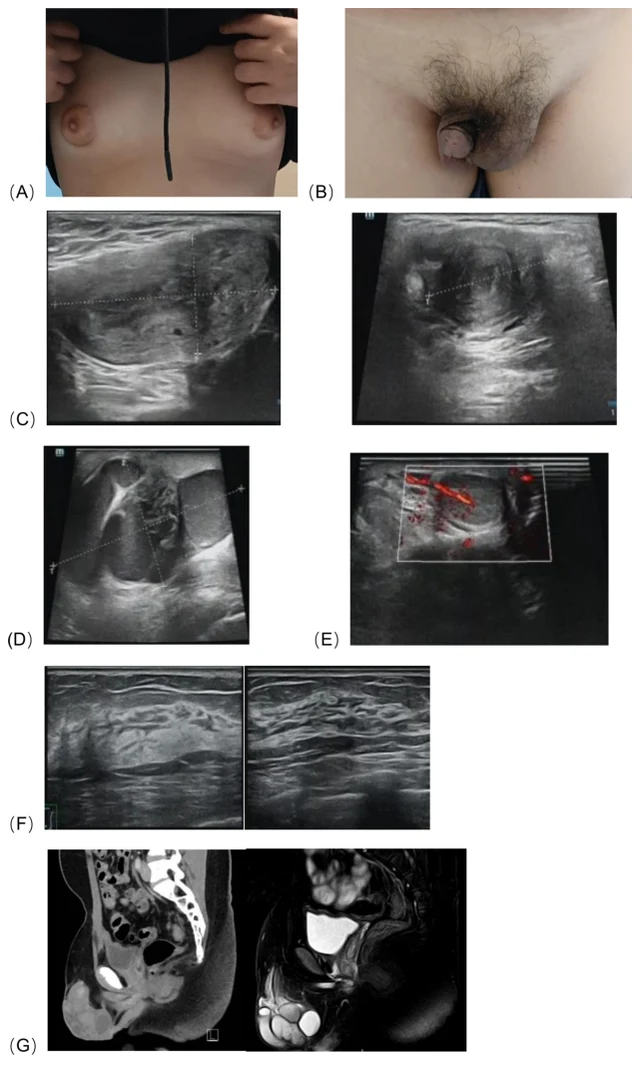
FIGURE 1 (A) Bilateral breast development, (B) The patient ’ s external genitalia, (C) Ultrasonography revealed an abnormal solid mass in the left inguinal region and scrotum, consistent with a uterus, measuring approximately 5.7×3.0×2.4 cm, with endometrial echo (thickness ~0.7 cm) and uterine cavity separation (~0.4 cm). (D) Surrounding the mass were tortuous, dilated tubular anechoic areas, suggestive of fallopian tubes, measuring approximately 7.1×4.2×6.1 cm. (E) The right scrotum contained testicular parenchyma, measuring 2.0×1.4×0.8 cm. (F) Breast ultrasound showed glandular tissue in both breasts, measuring 1.2 cm in anteroposterior diameter, with no detectable masses. (G) CT and MRI revealed a tubular soft tissue structure with a lumen, measuring approximately 4.8×1.5×1.1 cm, was detected between the bladder and rectum.

Deslize para a esquerda ou direita para ver as imagens deste caso

Abrir imagem original
CasoPublicação originalAtlasVer a legenda original em chinês
DSD Research Library Versão dos dados v1.4.0