Ir para o conteúdo
DSD Research Library Plataforma pública de pesquisa
Português
  • 简体中文
  • English
  • Français
  • Русский
  • Español
  • Português
Pesquisa Casos Investigação Atlas Publicações Educação Métodos

Voltar à galeria de casos

46,XY DSD (diferenças do desenvolvimento sexual) · 46,XY · DSD-C022

IMG460856b9db8b33 · 2892 × 3899
CasoPublicação originalAtlasAbrir imagem original
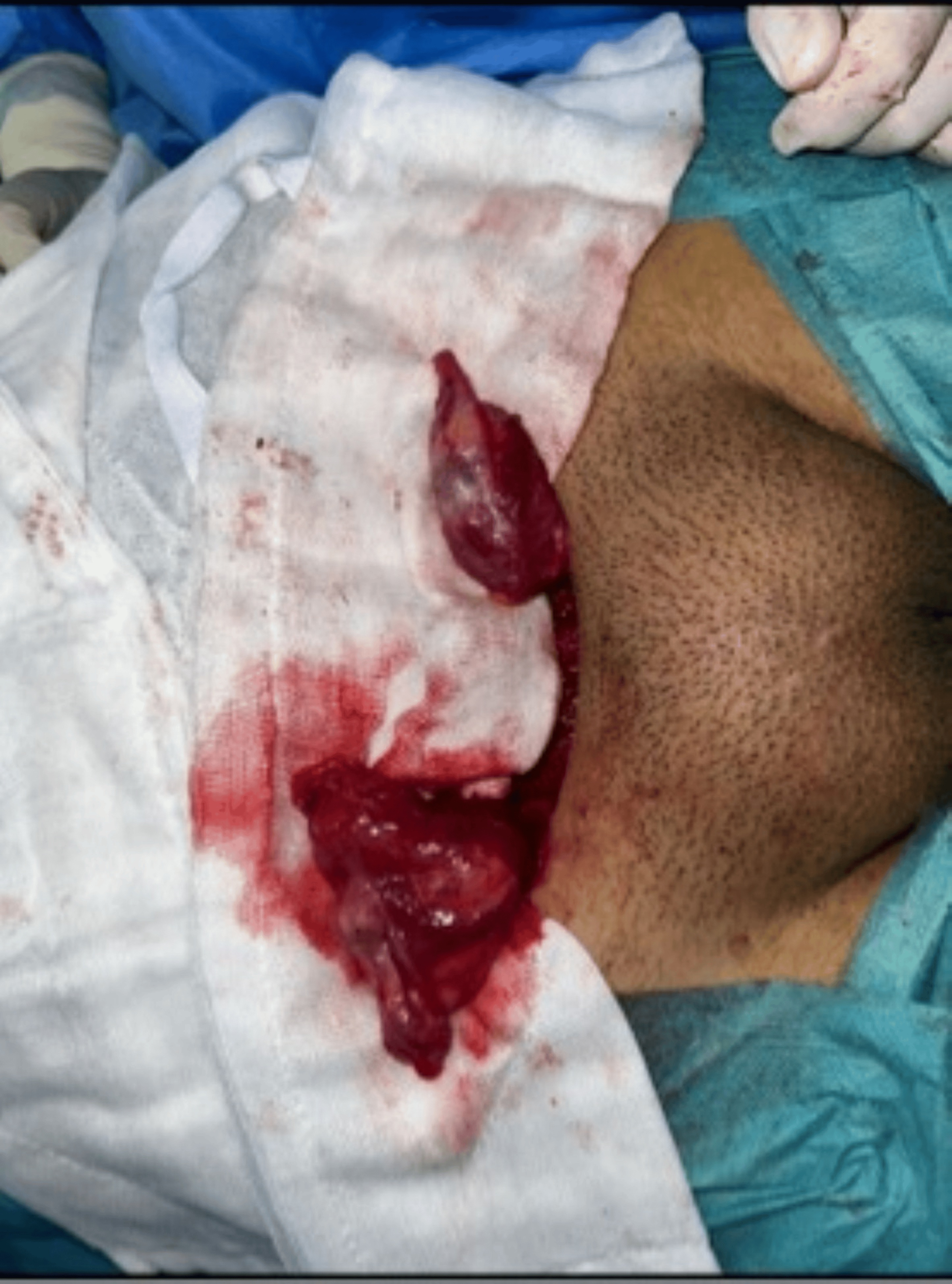
FIGURE 4: Intraoperative appearance of bilateral gonadal structures. Surgical exploration demonstrating dysgenetic gonadal tissue located in the inguinal region. Histopathological examination revealed dysgenetic testicular tissue with a limited number of medium-sized seminiferous tubules showing maturation arrest at the primary spermatocyte stage, sparse Leydig cells, and absence of ovarian parenchyma or stroma. These findings were consistent with dysgenetic testicular tissue. Management and follow-up Management was discussed in a multidisciplinary setting involving pediatric endocrinologists, pediatric surgeons, geneticists, and psychologists. Age-appropriate information was provided, and shared decision- making was conducted with the patient and her parents. The patient expressed a stable female gender identity. Bilateral orchiectomy was performed in accordance with the declared gender, with psychological support. Histological analysis of the resected gonads showed complete testicular atrophy without intratubular germ cell neoplasia or malignant features. Estrogen replacement therapy with 17β-estradiol was subsequently initiated, along with management of hyperandrogenism and regular endocrinological follow-up.
Legenda da figura

FIGURE 4: Intraoperative appearance of bilateral gonadal structures. Surgical exploration demonstrating dysgenetic gonadal tissue located in the inguinal region. Histopathological examination revealed dysgenetic testicular tissue with a limited number of medium-sized seminiferous tubules showing maturation arrest at the primary spermatocyte stage, sparse Leydig cells, and absence of ovarian parenchyma or stroma. These findings were consistent with dysgenetic testicular tissue. Management and follow-up Management was discussed in a multidisciplinary setting involving pediatric endocrinologists, pediatric surgeons, geneticists, and psychologists. Age-appropriate information was provided, and shared decision- making was conducted with the patient and her parents. The patient expressed a stable female gender identity. Bilateral orchiectomy was performed in accordance with the declared gender, with psychological support. Histological analysis of the resected gonads showed complete testicular atrophy without intratubular germ cell neoplasia or malignant features. Estrogen replacement therapy with 17β-estradiol was subsequently initiated, along with management of hyperandrogenism and regular endocrinological follow-up.

Imagens · 1
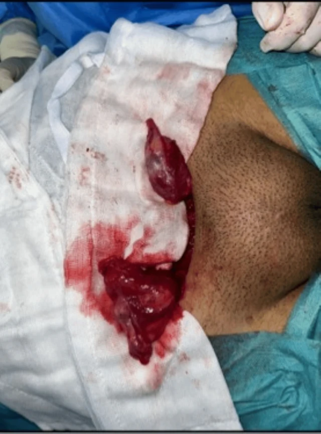
FIGURE 4: Intraoperative appearance of bilateral gonadal structures. Surgical exploration demonstrating dysgenetic gonadal tissue located in the inguinal region. Histopathological examination revealed dysgenetic testicular tissue with a limited number of medium-sized seminiferous tubules showing maturation arrest at the primary spermatocyte stage, sparse Leydig cells, and absence of ovarian parenchyma or stroma. These findings were consistent with dysgenetic testicular tissue. Management and follow-up Management was discussed in a multidisciplinary setting involving pediatric endocrinologists, pediatric surgeons, geneticists, and psychologists. Age-appropriate information was provided, and shared decision- making was conducted with the patient and her parents. The patient expressed a stable female gender identity. Bilateral orchiectomy was performed in accordance with the declared gender, with psychological support. Histological analysis of the resected gonads showed complete testicular atrophy without intratubular germ cell neoplasia or malignant features. Estrogen replacement therapy with 17β-estradiol was subsequently initiated, along with management of hyperandrogenism and regular endocrinological follow-up.

Deslize para a esquerda ou direita para ver as imagens deste caso

Abrir imagem original
CasoPublicação originalAtlasVer a legenda original em chinês
DSD Research Library Versão dos dados v1.4.0