Ir para o conteúdo
DSD Research Library Plataforma pública de pesquisa
Português
  • 简体中文
  • English
  • Français
  • Русский
  • Español
  • Português
Pesquisa Casos Investigação Atlas Publicações Educação Métodos

Voltar à galeria de casos

DSD 46,XX (seudohermafroditismo feminino, exposição intrauterina a andrógenos por adenoma do córtex suprarrenal materno). · 46,XX · DSD-C029

IMGcbb803a64a8e4f · 507 × 552
CasoPublicação originalAtlasAbrir imagem original
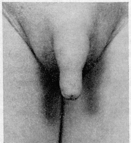
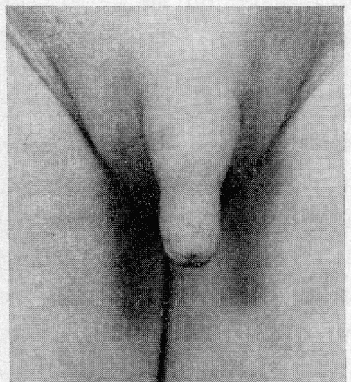
Fig. 2. Male external genitalia of the same girl. The karyotype was 46XX. Serum electrolytes were within normal limits. The results of the steroid studies are presented below in more detail. A voiding cysto-urethrogram showed no anomalies. At surgery, the right inguinal canal was opened and no gonad was found. On revision of the abdominal cavity, two bean-sized gonads, two Fallopian tubes, and a small uterus attached to the bladder were detected. No Wolffian structures were found. A biopsy was taken from each gonad. Histological examination revealed ovarian tissue in both biopsies and 42 per cent of the
Legenda da figura

Fig. 2. Male external genitalia of the same girl. The karyotype was 46XX. Serum electrolytes were within normal limits. The results of the steroid studies are presented below in more detail. A voiding cysto-urethrogram showed no anomalies. At surgery, the right inguinal canal was opened and no gonad was found. On revision of the abdominal cavity, two bean-sized gonads, two Fallopian tubes, and a small uterus attached to the bladder were detected. No Wolffian structures were found. A biopsy was taken from each gonad. Histological examination revealed ovarian tissue in both biopsies and 42 per cent of the

Imagens · 2
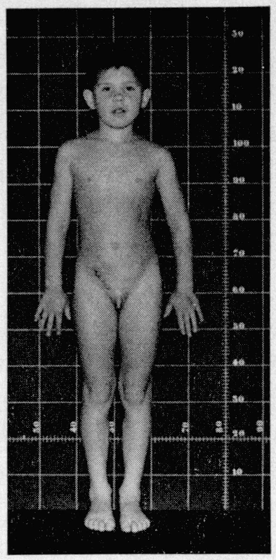
Fig. 1. 7 years old girl with female pseudohermaphroditism due to virilizing adenoma of the mother. Fig. 2. Male external genitalia of the same girl. The karyotype was 46XX. Serum electrolytes were within normal limits. The results of the steroid studies are presented below in more detail. A voiding cysto-urethrogram showed no anomalies. At surgery, the right inguinal canal was opened and no gonad was found. On revision of the abdominal cavity, two bean-sized gonads, two Fallopian tubes, and a small uterus attached to the bladder were detected. No Wolffian structures were found. A biopsy was taken from each gonad. Histological examination revealed ovarian tissue in both biopsies and 42 per cent of the

Deslize para a esquerda ou direita para ver as imagens deste caso

Abrir imagem original
CasoPublicação originalAtlasVer a legenda original em chinês
DSD Research Library Versão dos dados v1.4.0