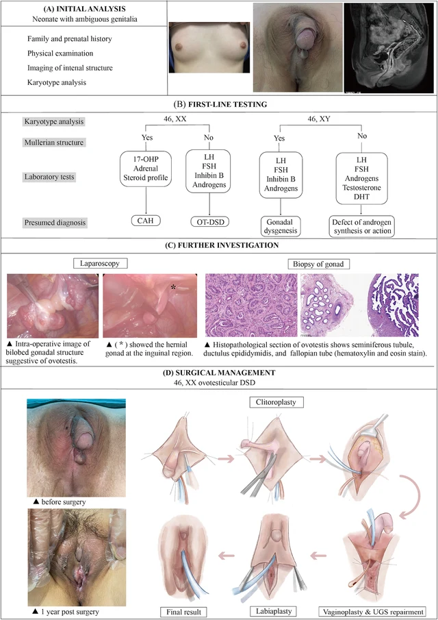
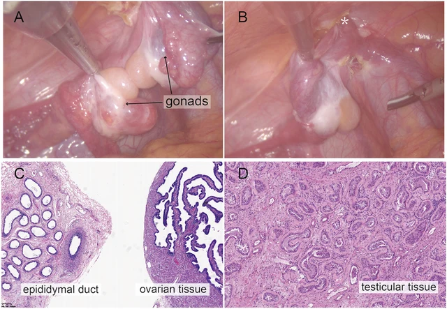
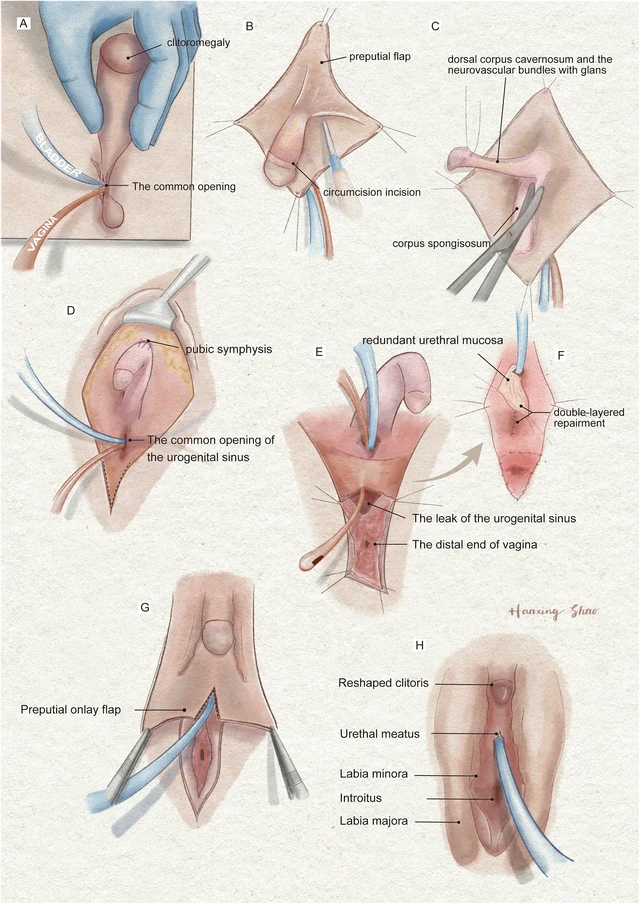
DSD ovotesticulaire 46,XX (OT-DSD). · 46,XX · DSD-C011
IMGe107aa5ed9f757 · 1725 × 1366
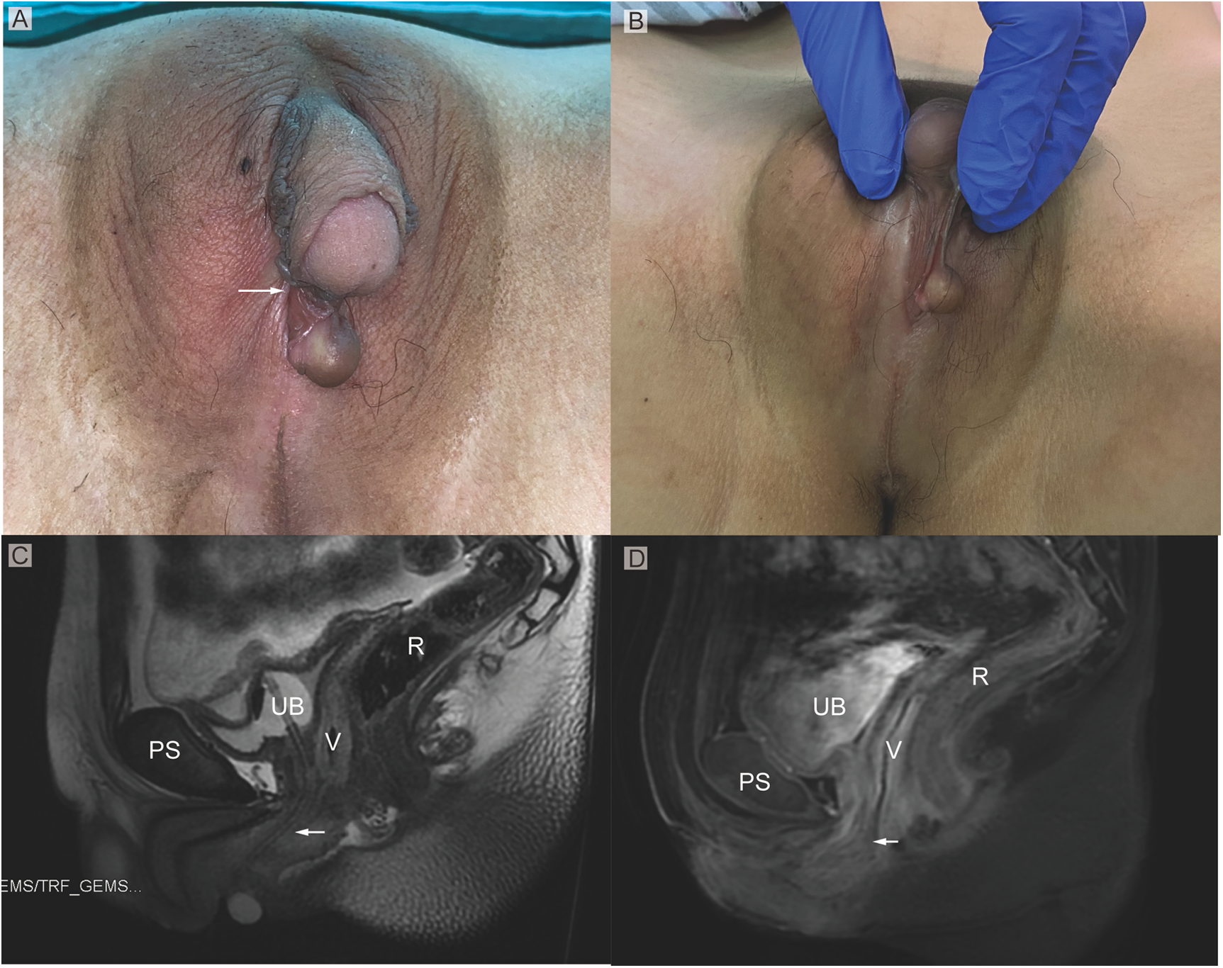
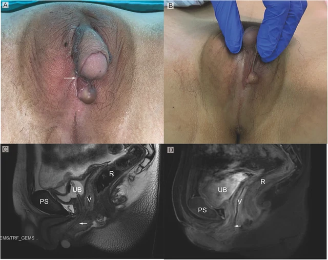
FIGURE 1 (A) Phenotypic appearance of genitalia before surgery. An enlarged clitoris, completely fused labia majora, and a movable painless mass at the right inguinal region was found. (B) An opening of the urogenital sinus was seen at the base of the clitoris. (C,D) Preoperative contrast-enhanced MRI showing well-defined urogenital sinus. Uterus was not detected. Sagittal MRI scan showing urinary bladder (UB), vagina (V), the common opening of the urogenital sinus (arrow), and rectum (R) The distance between the conference point to the superficial perineal pouch was below the level of the distal end of pubic symphysis (PS). Zhao et al. 10.3389/fsurg.2024.1502340 Frontiers in Surgery 02 frontiersin.org
Images · 4