Перейти к содержанию
DSD Research Library Открытая исследовательская платформа
Русский
  • 简体中文
  • English
  • Français
  • Русский
  • Español
  • Português
Поиск Регистр случаев Исследования Атлас Публикации Обучение Методы

Назад в галерею случаев

46,XX овотестис-ДСП (OT-DSD). · 46,XX · DSD-C011

IMGe107aa5ed9f757 · 1725 × 1366
СлучайПервоисточникАтласОткрыть исходное изображение
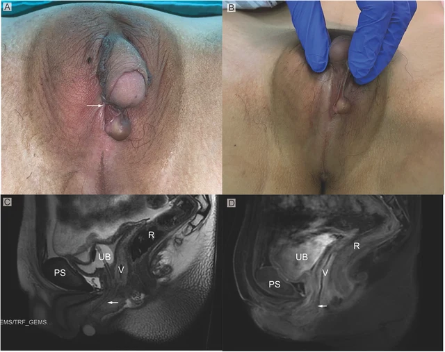
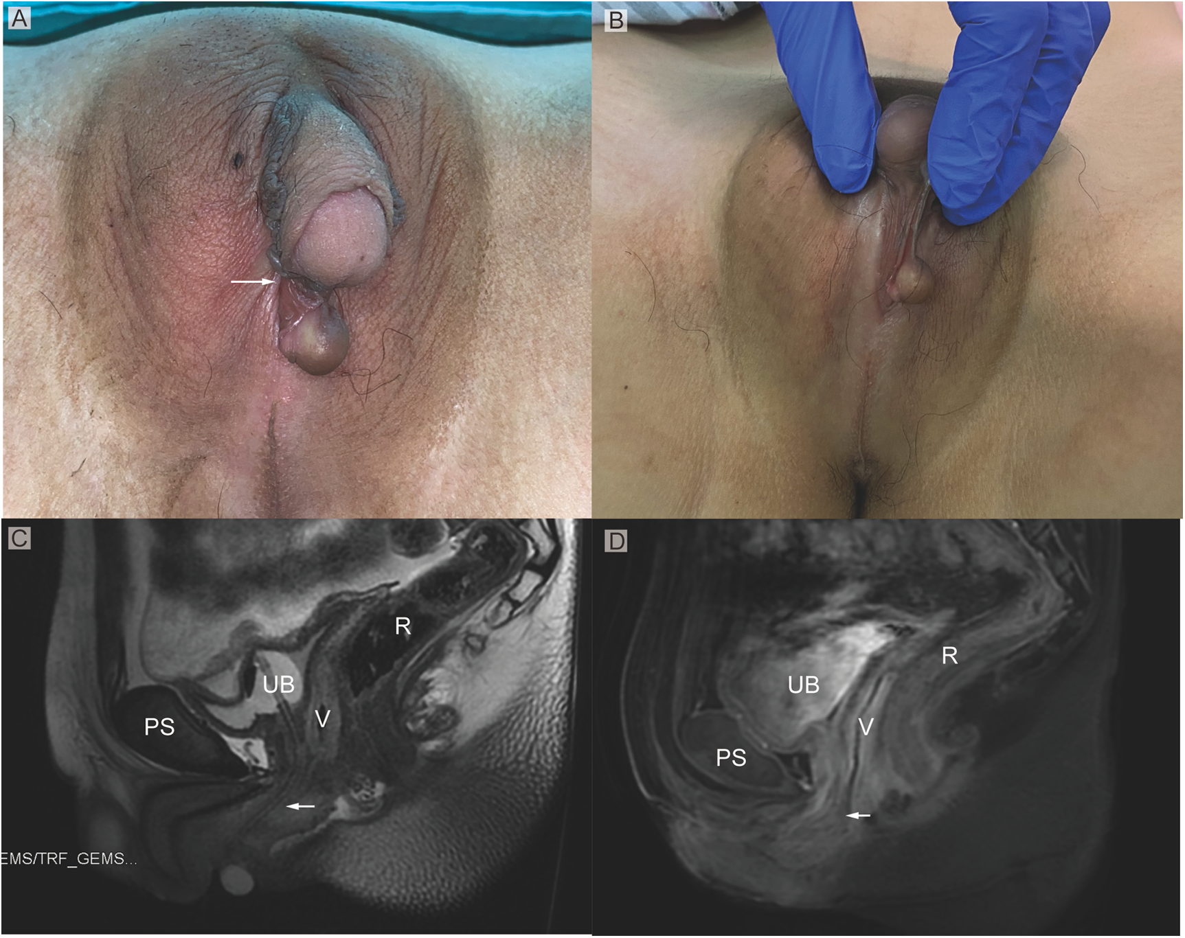
FIGURE 1 (A) Phenotypic appearance of genitalia before surgery. An enlarged clitoris, completely fused labia majora, and a movable painless mass at the right inguinal region was found. (B) An opening of the urogenital sinus was seen at the base of the clitoris. (C,D) Preoperative contrast-enhanced MRI showing well-defined urogenital sinus. Uterus was not detected. Sagittal MRI scan showing urinary bladder (UB), vagina (V), the common opening of the urogenital sinus (arrow), and rectum (R) The distance between the conference point to the superficial perineal pouch was below the level of the distal end of pubic symphysis (PS). Zhao et al. 10.3389/fsurg.2024.1502340 Frontiers in Surgery 02 frontiersin.org
Подпись к рисунку

FIGURE 1 (A) Phenotypic appearance of genitalia before surgery. An enlarged clitoris, completely fused labia majora, and a movable painless mass at the right inguinal region was found. (B) An opening of the urogenital sinus was seen at the base of the clitoris. (C,D) Preoperative contrast-enhanced MRI showing well-defined urogenital sinus. Uterus was not detected. Sagittal MRI scan showing urinary bladder (UB), vagina (V), the common opening of the urogenital sinus (arrow), and rectum (R) The distance between the conference point to the superficial perineal pouch was below the level of the distal end of pubic symphysis (PS). Zhao et al. 10.3389/fsurg.2024.1502340 Frontiers in Surgery 02 frontiersin.org

Изображения · 4
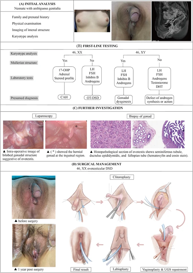
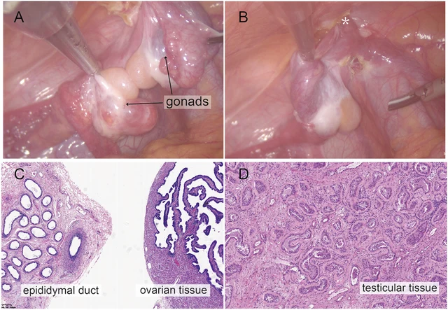
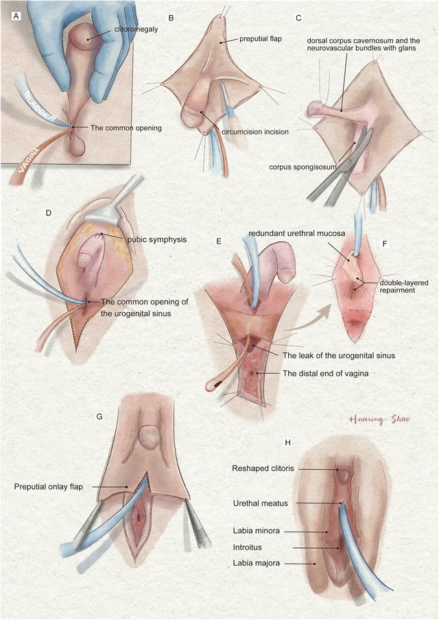
FIGURE 4 The illustration of the systematic diagnostic and treatment process for OT-DSD. The investigation process of OT-DSD can be summarized as initial assessment (A), first-line testing (B), and further investigation (C,D) showed the procedures of feminizing genitoplasty and pre- and 1-year post- operative pictures. The procedures included clitoroplasty, vaginoplasty and urogenital sinus repair, along with labiaplasty. 17, OHP, 17-alpha hydroxy progesterone; LH, Luteinizing hormone; FSH, follicle-stimulating hormone; DHT, dihydrotestosterone; CAH, congenital adrenal hyperplasia; OT-DSD, ovotesticular disorder of sex development. Zhao et al. 10.3389/fsurg.2024.1502340 Frontiers in Surgery 06 frontiersin.org FIGURE 2 Intraoperative findings from laparoscopic exploration. (A) Intra-operative image of heterogenous gonadal structure showed the testicular and ovarian fractions organized in an end-to-end fashion, suggesting the ovotestis. (B) The right gonad was found at the inner ring of inguinal canal (*). (C) A biopsy was taken from the right hernial gonad in order to rule out any dysgenesis or ovotesticular tissue. Histopathological examination of sample 1 revealed epididymal and ovarian tissue. (D) Histopathological examination of sample 2 showed testicular histology with seminiferous tubules. Zhao et al. 10.3389/fsurg.2024.1502340 Frontiers in Surgery 03 frontiersin.org FIGURE 1 (A) Phenotypic appearance of genitalia before surgery. An enlarged clitoris, completely fused labia majora, and a movable painless mass at the right inguinal region was found. (B) An opening of the urogenital sinus was seen at the base of the clitoris. (C,D) Preoperative contrast-enhanced MRI showing well-defined urogenital sinus. Uterus was not detected. Sagittal MRI scan showing urinary bladder (UB), vagina (V), the common opening of the urogenital sinus (arrow), and rectum (R) The distance between the conference point to the superficial perineal pouch was below the level of the distal end of pubic symphysis (PS). Zhao et al. 10.3389/fsurg.2024.1502340 Frontiers in Surgery 02 frontiersin.org FIGURE 3 The illustration of the surgical procedures of feminizing genitoplasty. (A) The vulva showed Prader 4 virilization. (B) A subcoronal circumferential incision was made to de-glove the clitoris over Buck’s fascia. (C) The corporal bodies were excised with preservation of the glans clitoris and neurovascular bundles. (D) The corpora cavernosa was folded and sutured to the pubic arch. (E) The orange urethral catheter entered from the orifice through the urogenital sinus to exit from the fistula. (F) The fistula was sutured and secured with the rotating urethral mucosa flap. The vagina was pull-down to the vulve and sutured to the skin. (G) The prepuce in an onlay fashion covers the area between the clitoris and the vestibule. (H) The final result of the procedure. Zhao et al. 10.3389/fsurg.2024.1502340 Frontiers in Surgery 05 frontiersin.org

Листайте влево или вправо, чтобы просмотреть изображения этого случая

Открыть исходное изображение
СлучайПервоисточникАтласИсходная подпись на китайском
DSD Research Library Версия данных v1.4.0