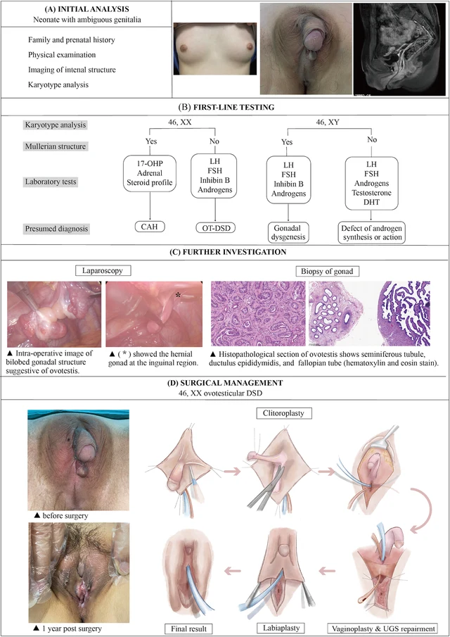
46,XX овотестис-ДСП (OT-DSD). · 46,XX · DSD-C011
IMGa96d981c000e5e · 1725 × 1194
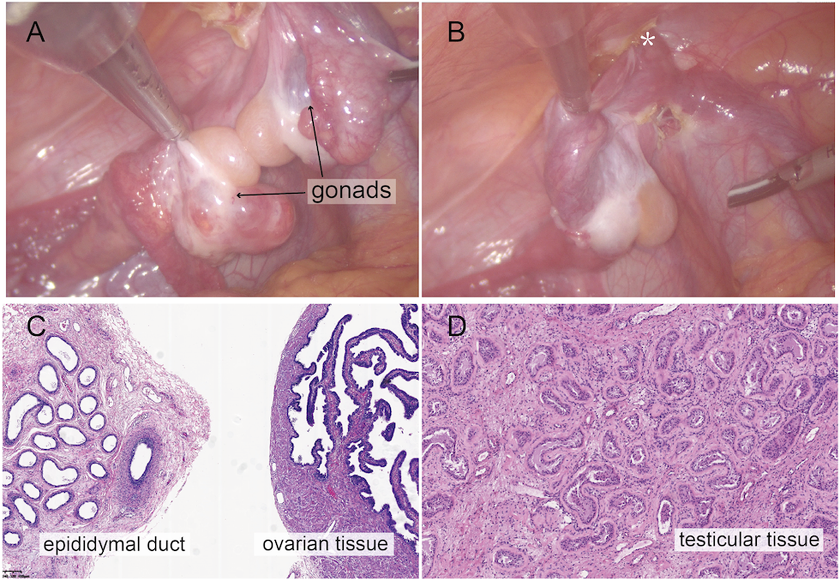
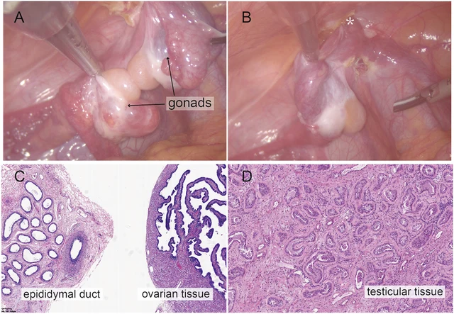
FIGURE 2 Intraoperative findings from laparoscopic exploration. (A) Intra-operative image of heterogenous gonadal structure showed the testicular and ovarian fractions organized in an end-to-end fashion, suggesting the ovotestis. (B) The right gonad was found at the inner ring of inguinal canal (*). (C) A biopsy was taken from the right hernial gonad in order to rule out any dysgenesis or ovotesticular tissue. Histopathological examination of sample 1 revealed epididymal and ovarian tissue. (D) Histopathological examination of sample 2 showed testicular histology with seminiferous tubules. Zhao et al. 10.3389/fsurg.2024.1502340 Frontiers in Surgery 03 frontiersin.org
Изображения · 4