Skip to content
DSD Research Library Public Research Platform
English
  • 简体中文
  • English
  • Français
  • Русский
  • Español
  • Português
Search Cases Research Atlas Papers Learn Methods

Back to the case gallery

Gonadal dysgenesis · 47,XYY;45,X/46,XY · DSD-C073

IMG130344a67b6c9c · 934 × 480
CaseSource publicationAtlasOpen original image
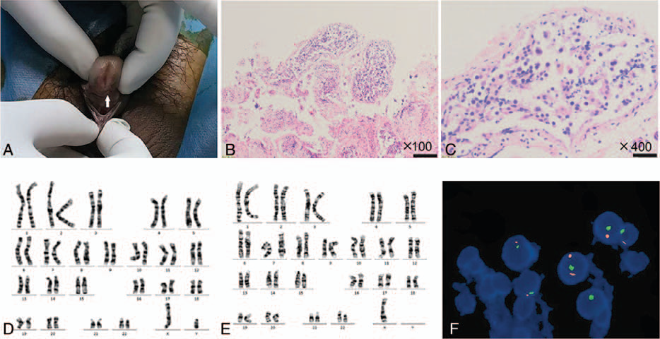
Figure 1. Diagnosis of 45,X/46,XY mosaicism. The patient?s penis is short. The urethral opening (arrow) is on the middle and anterior parts of the ventral side of the penis (A). H&E staining at 100? (B) and 400? (C) showed seminiferous tubule dysgenesis. Chromosomal analysis showed a 46,XY karyotype in (D) and 45,X in (E). FISH analysis revealed a 45,X/46,XY/47,XYY karyotype in testicular tissue (F).
Figure caption

Figure 1. Diagnosis of 45,X/46,XY mosaicism. The patient?s penis is short. The urethral opening (arrow) is on the middle and anterior parts of the ventral side of the penis (A). H&E staining at 100? (B) and 400? (C) showed seminiferous tubule dysgenesis. Chromosomal analysis showed a 46,XY karyotype in (D) and 45,X in (E). FISH analysis revealed a 45,X/46,XY/47,XYY karyotype in testicular tissue (F).

Images · 1
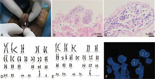
Figure 1. Diagnosis of 45,X/46,XY mosaicism. The patient?s penis is short. The urethral opening (arrow) is on the middle and anterior parts of the ventral side of the penis (A). H&E staining at 100? (B) and 400? (C) showed seminiferous tubule dysgenesis. Chromosomal analysis showed a 46,XY karyotype in (D) and 45,X in (E). FISH analysis revealed a 45,X/46,XY/47,XYY karyotype in testicular tissue (F).

Swipe left or right to browse this case’s images

Open original image
CaseSource publicationAtlasView original caption in Chinese
DSD Research Library Data version v1.4.0