跳至正文
DSD Research Library 国际化科研证据平台
简体中文
  • 简体中文
  • English
  • Français
  • Русский
  • Español
  • Português
全站检索 病例注册库 研究证据矩阵 知识图谱 文献数据库 医学教育 方法与引用

返回病例图库

性腺发育不良 · 47,XYY;45,X/46,XY · DSD-C073

IMG130344a67b6c9c · 934 × 480
病例来源论文知识图谱查看原图
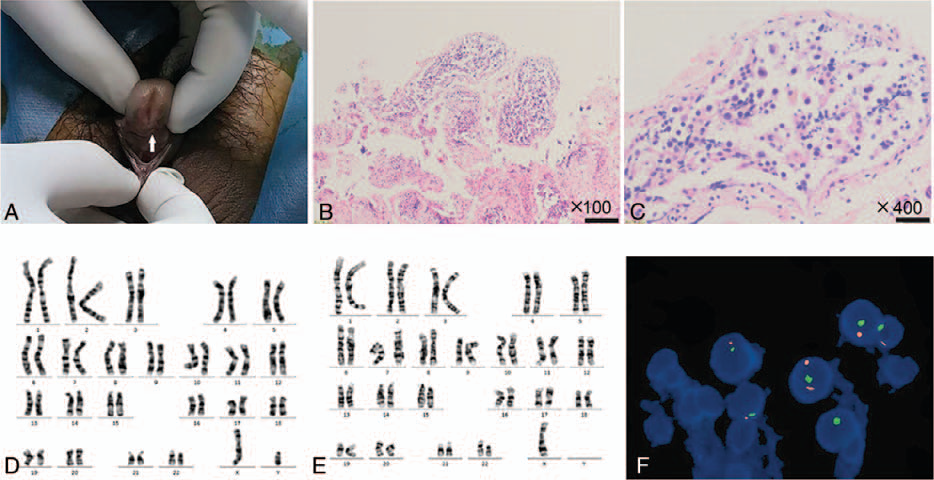
Figure 1. Diagnosis of 45,X/46,XY mosaicism. The patient’s penis is short. The urethral opening (arrow) is on the middle and anterior parts of the ventral side of the penis (A). H&E staining at 100× (B) and 400× (C) showed seminiferous tubule dysgenesis. Chromosomal analysis showed a 46,XY karyotype in (D) and 45,X in (E). FISH analysis revealed a 45,X/46,XY/47,XYY karyotype in testicular tissue (F).
图注

Figure 1. Diagnosis of 45,X/46,XY mosaicism. The patient’s penis is short. The urethral opening (arrow) is on the middle and anterior parts of the ventral side of the penis (A). H&E staining at 100× (B) and 400× (C) showed seminiferous tubule dysgenesis. Chromosomal analysis showed a 46,XY karyotype in (D) and 45,X in (E). FISH analysis revealed a 45,X/46,XY/47,XYY karyotype in testicular tissue (F).

配图 · 1
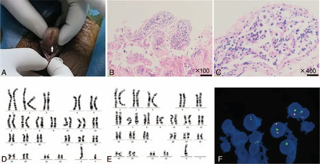
Figure 1. Diagnosis of 45,X/46,XY mosaicism. The patient’s penis is short. The urethral opening (arrow) is on the middle and anterior parts of the ventral side of the penis (A). H&E staining at 100× (B) and 400× (C) showed seminiferous tubule dysgenesis. Chromosomal analysis showed a 46,XY karyotype in (D) and 45,X in (E). FISH analysis revealed a 45,X/46,XY/47,XYY karyotype in testicular tissue (F).

左右滑动,浏览同病例配图

查看原图
病例来源论文知识图谱查看中文原始图注
DSD Research Library 数据版本 v1.4.0