Перейти к содержанию
DSD Research Library Открытая исследовательская платформа
Русский
  • 简体中文
  • English
  • Français
  • Русский
  • Español
  • Português
Поиск Регистр случаев Исследования Атлас Публикации Обучение Методы

Назад в галерею случаев

Дисгенезия гонад · 47,XYY;45,X/46,XY · DSD-C073

IMG130344a67b6c9c · 934 × 480
СлучайПервоисточникАтласОткрыть исходное изображение
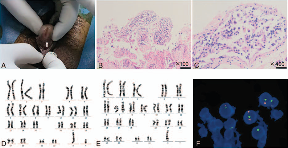
Figure 1. Diagnosis of 45,X/46,XY mosaicism. The patient?s penis is short. The urethral opening (arrow) is on the middle and anterior parts of the ventral side of the penis (A). H&E staining at 100? (B) and 400? (C) showed seminiferous tubule dysgenesis. Chromosomal analysis showed a 46,XY karyotype in (D) and 45,X in (E). FISH analysis revealed a 45,X/46,XY/47,XYY karyotype in testicular tissue (F).
Подпись к рисунку

Figure 1. Diagnosis of 45,X/46,XY mosaicism. The patient?s penis is short. The urethral opening (arrow) is on the middle and anterior parts of the ventral side of the penis (A). H&E staining at 100? (B) and 400? (C) showed seminiferous tubule dysgenesis. Chromosomal analysis showed a 46,XY karyotype in (D) and 45,X in (E). FISH analysis revealed a 45,X/46,XY/47,XYY karyotype in testicular tissue (F).

Изображения · 1
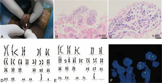
Figure 1. Diagnosis of 45,X/46,XY mosaicism. The patient?s penis is short. The urethral opening (arrow) is on the middle and anterior parts of the ventral side of the penis (A). H&E staining at 100? (B) and 400? (C) showed seminiferous tubule dysgenesis. Chromosomal analysis showed a 46,XY karyotype in (D) and 45,X in (E). FISH analysis revealed a 45,X/46,XY/47,XYY karyotype in testicular tissue (F).

Листайте влево или вправо, чтобы просмотреть изображения этого случая

Открыть исходное изображение
СлучайПервоисточникАтласИсходная подпись на китайском
DSD Research Library Версия данных v1.4.0