Skip to content
DSD Research Library Public Research Platform
English
  • 简体中文
  • English
  • Français
  • Русский
  • Español
  • Português
Search Cases Research Atlas Papers Learn Methods

Back to the case gallery

Other / unclassified DSD · 46,XX · DSD-C040

IMGd2c91d36eba0c3 · 843 × 698
CaseSource publicationAtlasOpen original image
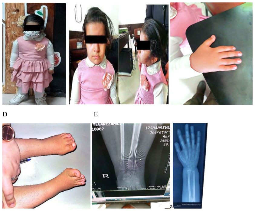
FIGURE 2 | (A) Full-body photograph at age 5, demonstrating disproportionate short stature (height: 88 cm, Z score − 4.4; upper-to-lower body segment ratio: 1.8). (B) Clinical image highlighting facial dysmorphism, including frontal bossing, mild hypertropia of the right eye, low-set ears, and a short neck, consistent with IMAGe syndrome. (C) Photograph of the right hand showing a shortened index finger. (D) Image of the feet demonstrating bilateral clinodactyly of the fourth toes, bending toward the third toes. (E) Radiographs of wrists and hips revealing osteonecrosis, metaphyseal dysplasia, and frayed bone edges.
Figure caption

FIGURE 2 | (A) Full-body photograph at age 5, demonstrating disproportionate short stature (height: 88 cm, Z score − 4.4; upper-to-lower body segment ratio: 1.8). (B) Clinical image highlighting facial dysmorphism, including frontal bossing, mild hypertropia of the right eye, low-set ears, and a short neck, consistent with IMAGe syndrome. (C) Photograph of the right hand showing a shortened index finger. (D) Image of the feet demonstrating bilateral clinodactyly of the fourth toes, bending toward the third toes. (E) Radiographs of wrists and hips revealing osteonecrosis, metaphyseal dysplasia, and frayed bone edges.

Images · 1
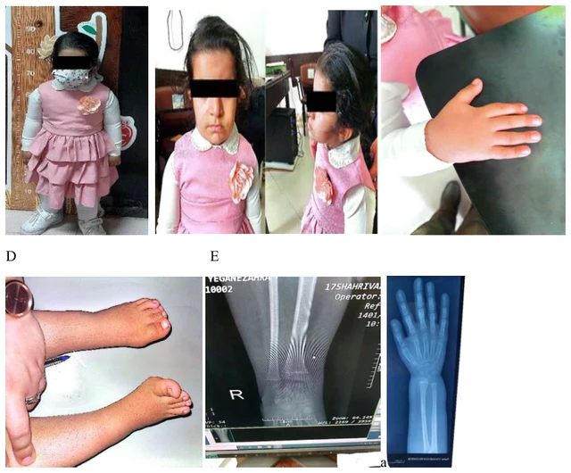
FIGURE 2 | (A) Full-body photograph at age 5, demonstrating disproportionate short stature (height: 88 cm, Z score − 4.4; upper-to-lower body segment ratio: 1.8). (B) Clinical image highlighting facial dysmorphism, including frontal bossing, mild hypertropia of the right eye, low-set ears, and a short neck, consistent with IMAGe syndrome. (C) Photograph of the right hand showing a shortened index finger. (D) Image of the feet demonstrating bilateral clinodactyly of the fourth toes, bending toward the third toes. (E) Radiographs of wrists and hips revealing osteonecrosis, metaphyseal dysplasia, and frayed bone edges.

Swipe left or right to browse this case’s images

Open original image
CaseSource publicationAtlasView original caption in Chinese
DSD Research Library Data version v1.4.0