Ir al contenido
DSD Research Library Plataforma pública de investigación
Español
  • 简体中文
  • English
  • Français
  • Русский
  • Español
  • Português
Búsqueda Casos Investigación Atlas Publicaciones Educación Métodos

Volver a la galería de casos

Otro/DSD no clasificado · 46,XX · DSD-C040

IMGd2c91d36eba0c3 · 843 × 698
CasoPublicación originalAtlasAbrir imagen original
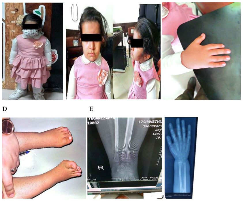
FIGURE 2 | (A) Full-body photograph at age 5, demonstrating disproportionate short stature (height: 88 cm, Z score − 4.4; upper-to-lower body segment ratio: 1.8). (B) Clinical image highlighting facial dysmorphism, including frontal bossing, mild hypertropia of the right eye, low-set ears, and a short neck, consistent with IMAGe syndrome. (C) Photograph of the right hand showing a shortened index finger. (D) Image of the feet demonstrating bilateral clinodactyly of the fourth toes, bending toward the third toes. (E) Radiographs of wrists and hips revealing osteonecrosis, metaphyseal dysplasia, and frayed bone edges.
Pie de figura

FIGURE 2 | (A) Full-body photograph at age 5, demonstrating disproportionate short stature (height: 88 cm, Z score − 4.4; upper-to-lower body segment ratio: 1.8). (B) Clinical image highlighting facial dysmorphism, including frontal bossing, mild hypertropia of the right eye, low-set ears, and a short neck, consistent with IMAGe syndrome. (C) Photograph of the right hand showing a shortened index finger. (D) Image of the feet demonstrating bilateral clinodactyly of the fourth toes, bending toward the third toes. (E) Radiographs of wrists and hips revealing osteonecrosis, metaphyseal dysplasia, and frayed bone edges.

Imágenes · 1
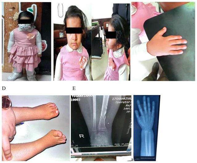
FIGURE 2 | (A) Full-body photograph at age 5, demonstrating disproportionate short stature (height: 88 cm, Z score − 4.4; upper-to-lower body segment ratio: 1.8). (B) Clinical image highlighting facial dysmorphism, including frontal bossing, mild hypertropia of the right eye, low-set ears, and a short neck, consistent with IMAGe syndrome. (C) Photograph of the right hand showing a shortened index finger. (D) Image of the feet demonstrating bilateral clinodactyly of the fourth toes, bending toward the third toes. (E) Radiographs of wrists and hips revealing osteonecrosis, metaphyseal dysplasia, and frayed bone edges.

Desliza a izquierda o derecha para ver las imágenes de este caso

Abrir imagen original
CasoPublicación originalAtlasVer el pie original en chino
DSD Research Library Versión de los datos v1.4.0