Перейти к содержанию
DSD Research Library Открытая исследовательская платформа
Русский
  • 简体中文
  • English
  • Français
  • Русский
  • Español
  • Português
Поиск Регистр случаев Исследования Атлас Публикации Обучение Методы

Назад в галерею случаев

Другие/неклассифицированные DSD · 46,XX · DSD-C040

IMGd2c91d36eba0c3 · 843 × 698
СлучайПервоисточникАтласОткрыть исходное изображение
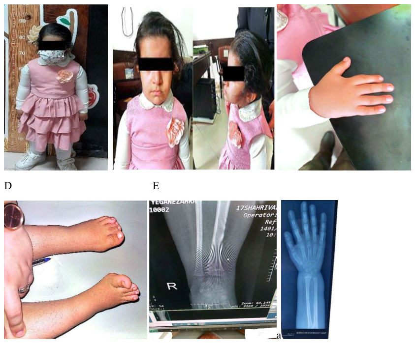
FIGURE 2 | (A) Full-body photograph at age 5, demonstrating disproportionate short stature (height: 88 cm, Z score − 4.4; upper-to-lower body segment ratio: 1.8). (B) Clinical image highlighting facial dysmorphism, including frontal bossing, mild hypertropia of the right eye, low-set ears, and a short neck, consistent with IMAGe syndrome. (C) Photograph of the right hand showing a shortened index finger. (D) Image of the feet demonstrating bilateral clinodactyly of the fourth toes, bending toward the third toes. (E) Radiographs of wrists and hips revealing osteonecrosis, metaphyseal dysplasia, and frayed bone edges.
Подпись к рисунку

FIGURE 2 | (A) Full-body photograph at age 5, demonstrating disproportionate short stature (height: 88 cm, Z score − 4.4; upper-to-lower body segment ratio: 1.8). (B) Clinical image highlighting facial dysmorphism, including frontal bossing, mild hypertropia of the right eye, low-set ears, and a short neck, consistent with IMAGe syndrome. (C) Photograph of the right hand showing a shortened index finger. (D) Image of the feet demonstrating bilateral clinodactyly of the fourth toes, bending toward the third toes. (E) Radiographs of wrists and hips revealing osteonecrosis, metaphyseal dysplasia, and frayed bone edges.

Изображения · 1
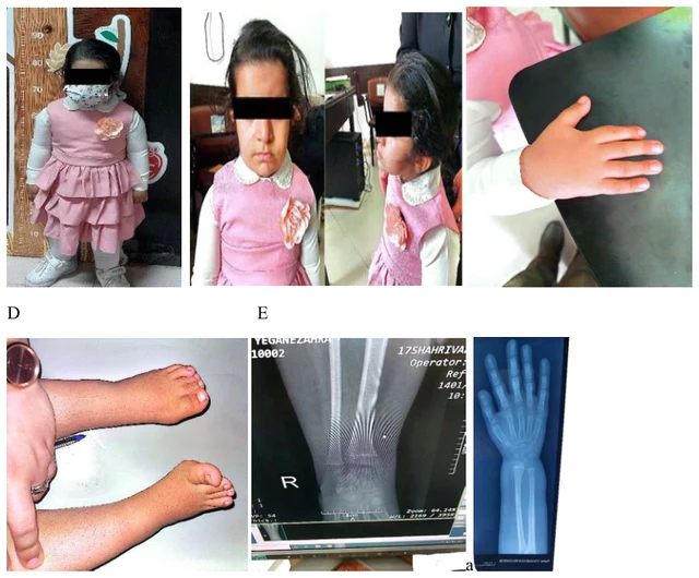
FIGURE 2 | (A) Full-body photograph at age 5, demonstrating disproportionate short stature (height: 88 cm, Z score − 4.4; upper-to-lower body segment ratio: 1.8). (B) Clinical image highlighting facial dysmorphism, including frontal bossing, mild hypertropia of the right eye, low-set ears, and a short neck, consistent with IMAGe syndrome. (C) Photograph of the right hand showing a shortened index finger. (D) Image of the feet demonstrating bilateral clinodactyly of the fourth toes, bending toward the third toes. (E) Radiographs of wrists and hips revealing osteonecrosis, metaphyseal dysplasia, and frayed bone edges.

Листайте влево или вправо, чтобы просмотреть изображения этого случая

Открыть исходное изображение
СлучайПервоисточникАтласИсходная подпись на китайском
DSD Research Library Версия данных v1.4.0