Aller au contenu
DSD Research Library Plateforme publique de recherche
Français
  • 简体中文
  • English
  • Français
  • Русский
  • Español
  • Português
Recherche Cas cliniques Recherche Atlas Publications Formation Méthodes

Retour à la galerie de cas

Autre DSD/non classé · 46,XY · DSD-C076

IMG3ffcc77eff2068 · 795 × 710
CasPublication sourceAtlasOuvrir l’image originale
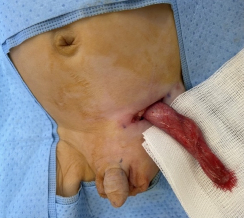
Fig. 3. Intraoperative clinical photographs of the left inguinoscrotal exploration in an 8-week-old phenotypical male with likely VSC: (a) Visible left inguinoscrotal swelling secondary to the indirect inguinal hernia and an empty right hemiscrotum. Normal appearing external male genitalia (b) Delivery of the left inguinal sac through the inguinal incision containing the two gonads identified on US imaging (c) Opening of the left patent processus vaginalis revealing two gonads with the accompanying uterus and fallopian tubes. These findings are most consistent with the diagnosis of TTE, which is one of the three main clinical presentations of PMDS.
Légende

Fig. 3. Intraoperative clinical photographs of the left inguinoscrotal exploration in an 8-week-old phenotypical male with likely VSC: (a) Visible left inguinoscrotal swelling secondary to the indirect inguinal hernia and an empty right hemiscrotum. Normal appearing external male genitalia (b) Delivery of the left inguinal sac through the inguinal incision containing the two gonads identified on US imaging (c) Opening of the left patent processus vaginalis revealing two gonads with the accompanying uterus and fallopian tubes. These findings are most consistent with the diagnosis of TTE, which is one of the three main clinical presentations of PMDS.

Images · 1
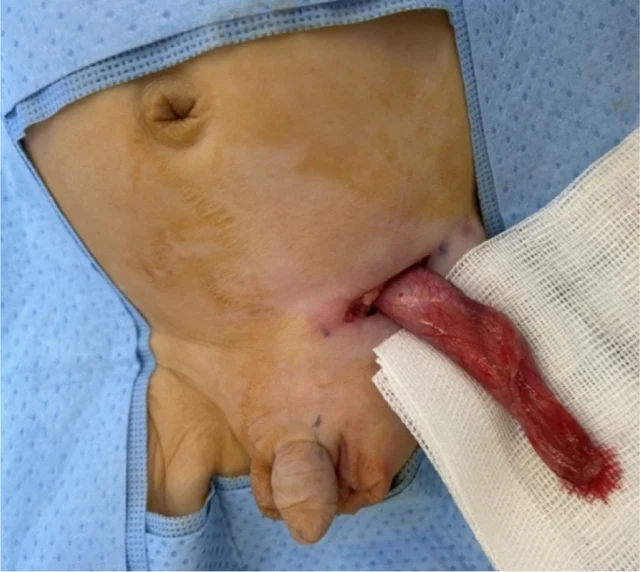
Fig. 3. Intraoperative clinical photographs of the left inguinoscrotal exploration in an 8-week-old phenotypical male with likely VSC: (a) Visible left inguinoscrotal swelling secondary to the indirect inguinal hernia and an empty right hemiscrotum. Normal appearing external male genitalia (b) Delivery of the left inguinal sac through the inguinal incision containing the two gonads identified on US imaging (c) Opening of the left patent processus vaginalis revealing two gonads with the accompanying uterus and fallopian tubes. These findings are most consistent with the diagnosis of TTE, which is one of the three main clinical presentations of PMDS.

Balayez à gauche ou à droite pour voir les images de ce cas

Ouvrir l’image originale
CasPublication sourceAtlasVoir la légende originale en chinois
DSD Research Library Version des données v1.4.0