Outro DSD / DSD não classificado · 46,XY · DSD-C076
IMG3ffcc77eff2068 · 795 × 710
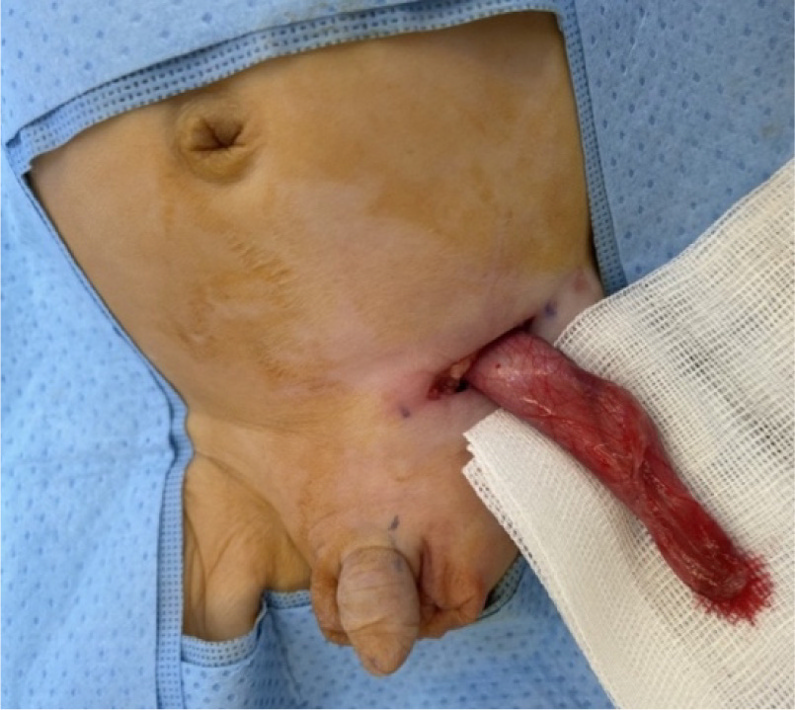
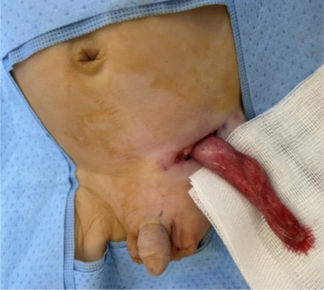
Fig. 3. Intraoperative clinical photographs of the left inguinoscrotal exploration in an 8-week-old phenotypical male with likely VSC: (a) Visible left inguinoscrotal swelling secondary to the indirect inguinal hernia and an empty right hemiscrotum. Normal appearing external male genitalia (b) Delivery of the left inguinal sac through the inguinal incision containing the two gonads identified on US imaging (c) Opening of the left patent processus vaginalis revealing two gonads with the accompanying uterus and fallopian tubes. These findings are most consistent with the diagnosis of TTE, which is one of the three main clinical presentations of PMDS.
Imagens · 1