Перейти к содержанию
DSD Research Library Открытая исследовательская платформа
Русский
  • 简体中文
  • English
  • Français
  • Русский
  • Español
  • Português
Поиск Регистр случаев Исследования Атлас Публикации Обучение Методы

Назад в галерею случаев

Другой/недифференцированный DSD · 46,XY · DSD-C076

IMG3ffcc77eff2068 · 795 × 710
СлучайПервоисточникАтласОткрыть исходное изображение
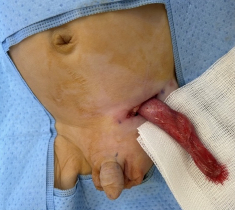
Fig. 3. Intraoperative clinical photographs of the left inguinoscrotal exploration in an 8-week-old phenotypical male with likely VSC: (a) Visible left inguinoscrotal swelling secondary to the indirect inguinal hernia and an empty right hemiscrotum. Normal appearing external male genitalia (b) Delivery of the left inguinal sac through the inguinal incision containing the two gonads identified on US imaging (c) Opening of the left patent processus vaginalis revealing two gonads with the accompanying uterus and fallopian tubes. These findings are most consistent with the diagnosis of TTE, which is one of the three main clinical presentations of PMDS.
Подпись к рисунку

Fig. 3. Intraoperative clinical photographs of the left inguinoscrotal exploration in an 8-week-old phenotypical male with likely VSC: (a) Visible left inguinoscrotal swelling secondary to the indirect inguinal hernia and an empty right hemiscrotum. Normal appearing external male genitalia (b) Delivery of the left inguinal sac through the inguinal incision containing the two gonads identified on US imaging (c) Opening of the left patent processus vaginalis revealing two gonads with the accompanying uterus and fallopian tubes. These findings are most consistent with the diagnosis of TTE, which is one of the three main clinical presentations of PMDS.

Изображения · 1
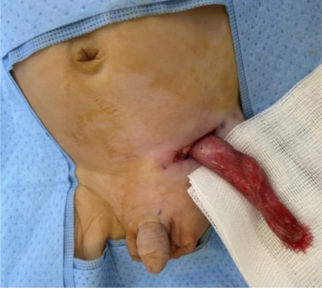
Fig. 3. Intraoperative clinical photographs of the left inguinoscrotal exploration in an 8-week-old phenotypical male with likely VSC: (a) Visible left inguinoscrotal swelling secondary to the indirect inguinal hernia and an empty right hemiscrotum. Normal appearing external male genitalia (b) Delivery of the left inguinal sac through the inguinal incision containing the two gonads identified on US imaging (c) Opening of the left patent processus vaginalis revealing two gonads with the accompanying uterus and fallopian tubes. These findings are most consistent with the diagnosis of TTE, which is one of the three main clinical presentations of PMDS.

Листайте влево или вправо, чтобы просмотреть изображения этого случая

Открыть исходное изображение
СлучайПервоисточникАтласИсходная подпись на китайском
DSD Research Library Версия данных v1.4.0