Ir al contenido
DSD Research Library Plataforma pública de investigación
Español
  • 简体中文
  • English
  • Français
  • Русский
  • Español
  • Português
Búsqueda Casos Investigación Atlas Publicaciones Educación Métodos

Galería de casos

Explora los casos e imágenes de la colección original. Busca por afección, gen, cariotipo o fenotipo y consulta la publicación original.

280 Casos con imágenes571 Imágenes

Registros de casosGalería de casos 280
Todos

Selección actual: 280 Casos con imágenes · 571 Imágenes

Selecciona una imagen para ampliarla. Las imágenes originales conservan el archivo fuente completo.

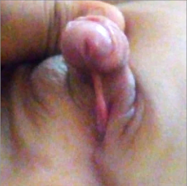
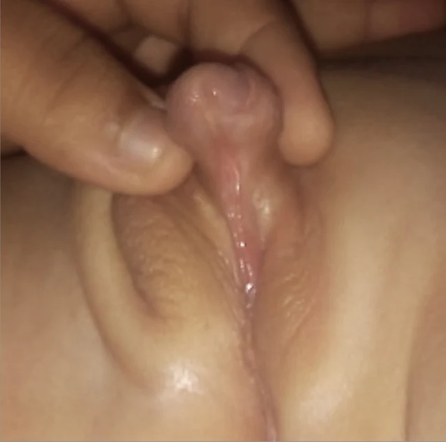
Imagen clínica · DSD-C01 3 Imágenes
Imagen clínica · DSD-C01 Imagen clínica · DSD-C01

DSD-C01

DSD ovotesticular · 46,XX · DSD-C01

46,XX · HPO:0000023, HPO:0000047, HPO:0000062, Inguinal hernia (HP:0000023), Ambiguous genitalia (HP:0000062), Hypospadias (HP:0000047)

Ovotestículo bipolar derecho, con tejido ovárico y testicular de aproximadamente 50% cada uno; la anatomía patológica no mostró tumor y la puntuación de potencial fértil del tejido testicular fue de 0.

CasoPublicación originalAtlas
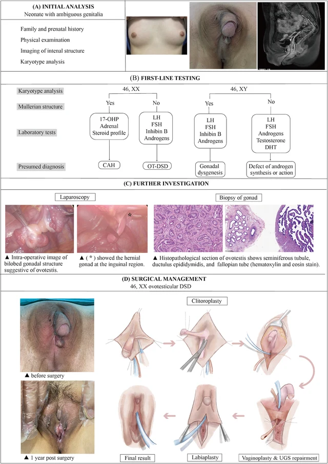
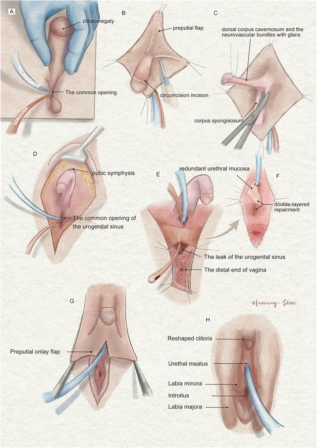
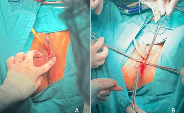
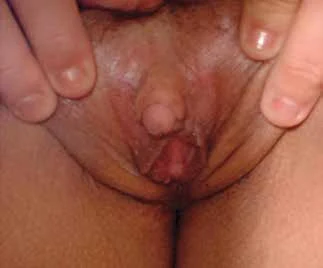
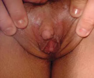
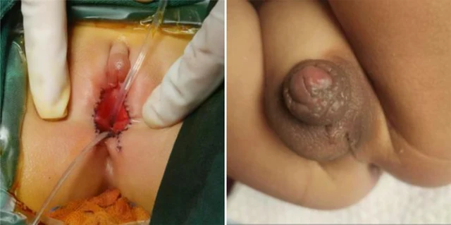
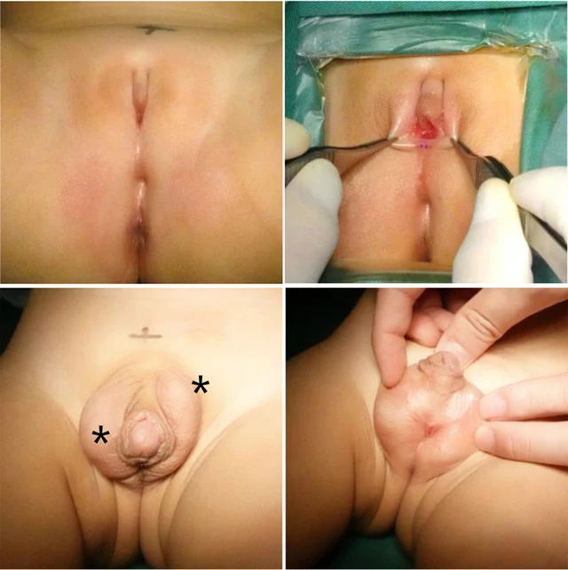
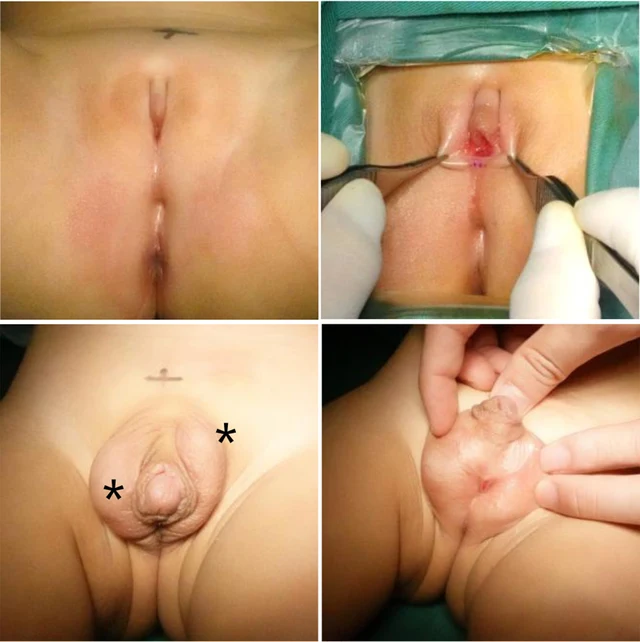
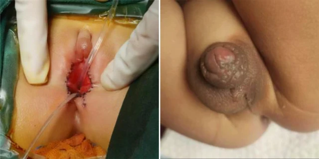
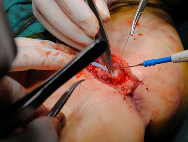
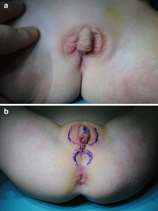
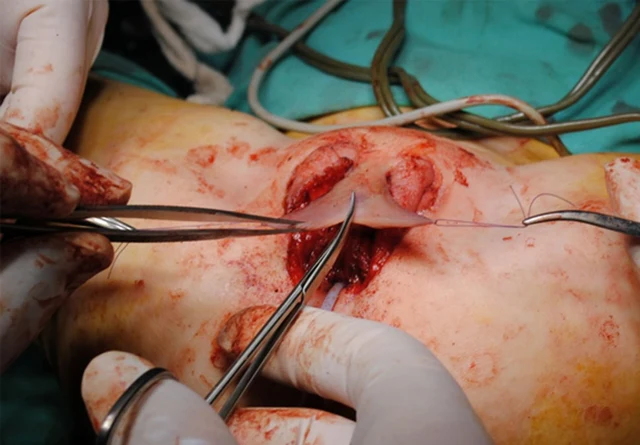
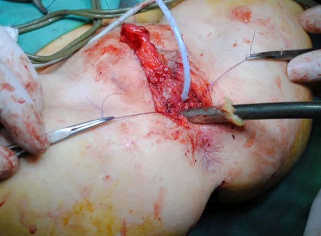
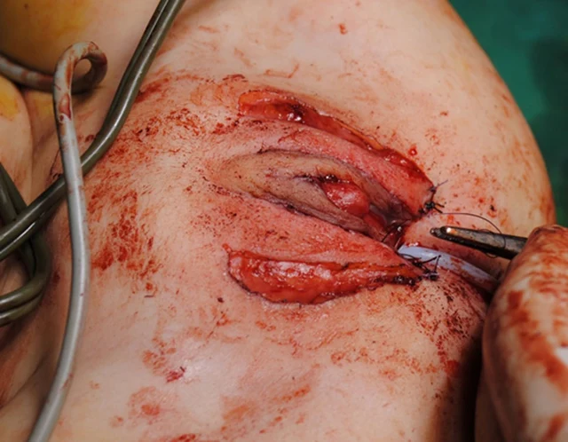
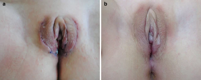
FIGURE 4 The illustration of the systematic diagnostic and treatment process for OT-DSD. The investigation process of OT-DSD can be summarized as initial assessment (A), first-line testing (B), and further investigation (C,D) showed the procedures of feminizing genitoplasty and pre- and 1-year post- operative pictures. The procedures included clitoroplasty, vaginoplasty and urogenital sinus repair, along with labiaplasty. 17, OHP, 17-alpha hydroxy progesterone; LH, Luteinizing hormone; FSH, follicle-stimulating hormone; DHT, dihydrotestosterone; CAH, congenital adrenal hyperplasia; OT-DSD, ovotesticular disorder of sex development. Zhao et al. 10.3389/fsurg.2024.1502340 Frontiers in Surgery 06 frontiersin.org 4 Imágenes
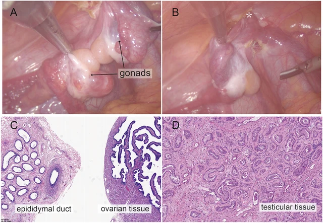
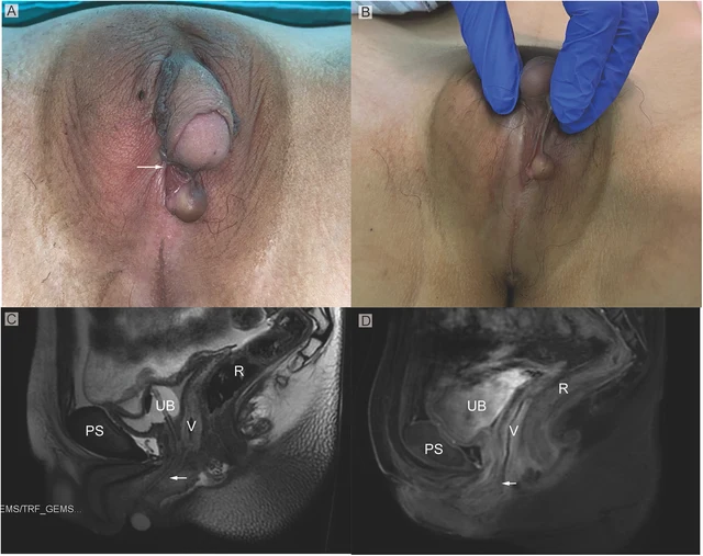
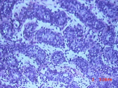
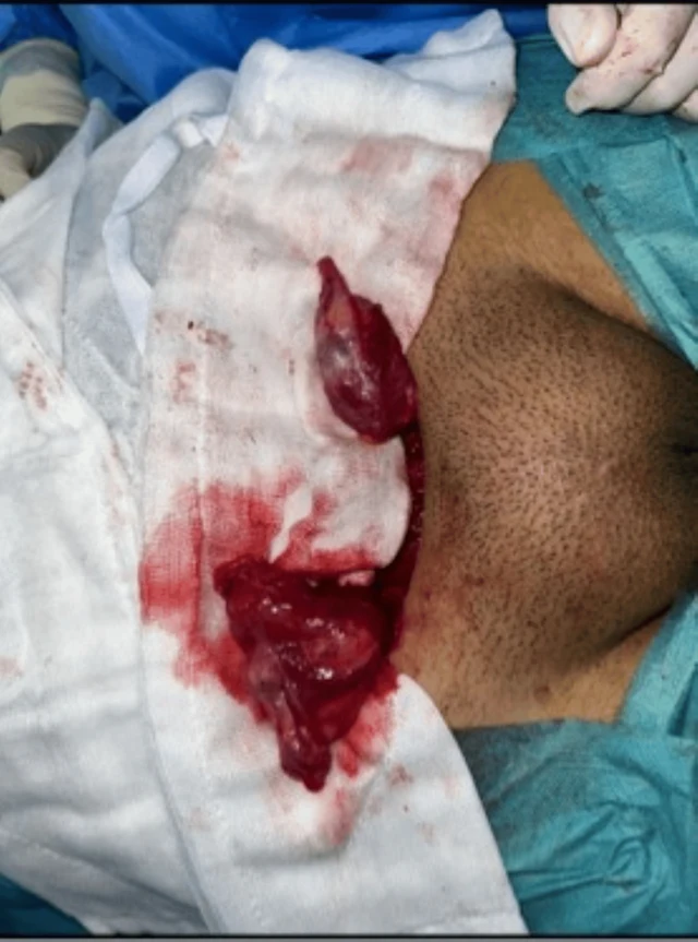
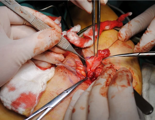
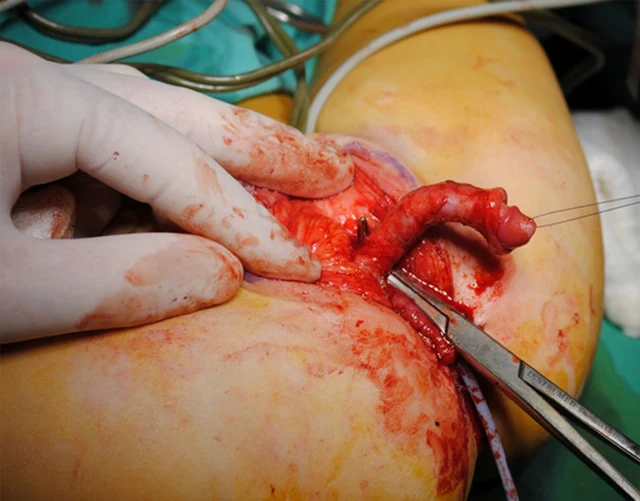
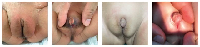
FIGURE 2 Intraoperative findings from laparoscopic exploration. (A) Intra-operative image of heterogenous gonadal structure showed the testicular and ovarian fractions organized in an end-to-end fashion, suggesting the ovotestis. (B) The right gonad was found at the inner ring of inguinal canal (*). (C) A biopsy was taken from the right hernial gonad in order to rule out any dysgenesis or ovotesticular tissue. Histopathological examination of sample 1 revealed epididymal and ovarian tissue. (D) Histopathological examination of sample 2 showed testicular histology with seminiferous tubules. Zhao et al. 10.3389/fsurg.2024.1502340 Frontiers in Surgery 03 frontiersin.org FIGURE 1 (A) Phenotypic appearance of genitalia before surgery. An enlarged clitoris, completely fused labia majora, and a movable painless mass at the right inguinal region was found. (B) An opening of the urogenital sinus was seen at the base of the clitoris. (C,D) Preoperative contrast-enhanced MRI showing well-defined urogenital sinus. Uterus was not detected. Sagittal MRI scan showing urinary bladder (UB), vagina (V), the common opening of the urogenital sinus (arrow), and rectum (R) The distance between the conference point to the superficial perineal pouch was below the level of the distal end of pubic symphysis (PS). Zhao et al. 10.3389/fsurg.2024.1502340 Frontiers in Surgery 02 frontiersin.org FIGURE 3 The illustration of the surgical procedures of feminizing genitoplasty. (A) The vulva showed Prader 4 virilization. (B) A subcoronal circumferential incision was made to de-glove the clitoris over Buck’s fascia. (C) The corporal bodies were excised with preservation of the glans clitoris and neurovascular bundles. (D) The corpora cavernosa was folded and sutured to the pubic arch. (E) The orange urethral catheter entered from the orifice through the urogenital sinus to exit from the fistula. (F) The fistula was sutured and secured with the rotating urethral mucosa flap. The vagina was pull-down to the vulve and sutured to the skin. (G) The prepuce in an onlay fashion covers the area between the clitoris and the vestibule. (H) The final result of the procedure. Zhao et al. 10.3389/fsurg.2024.1502340 Frontiers in Surgery 05 frontiersin.org

DSD-C011

46,XX DSD ovotesticular (OT-DSD). · 46,XX · DSD-C011

46,XX · HPO:0000047, Hypospadias (HP:0000047)

Estudios diagnósticos adicionales: la uretroscopia confirmó los hallazgos de la RM. Consulta multidisciplinaria de especialistas: reunión multidisciplinaria (MDT) centrada en el diagnóstico y la decisión quirúrgica, con participación de cirugía plástica, endocrinología, ginecología, urología, ginecología, psicología y trabajo social.

CasoPublicación originalAtlas
FIGURE 1 Genitalia abnormalities (a) & (b) External genitalia at 3 weeks. (c) Genitocystography. (d) Testicular biopsy a b c d 1 Imágenes

DSD-C012

46,XY DSD (disgenesia gonadal, deleción intersticial 8p23.1). · 46,XY · DSD-C012

AR, GATA4, SRD5A2, WT1 · 46,XY · HPO:0000028, HPO:0000047, HPO:0000054, Micropenis (HP:0000054), Hypospadias (HP:0000047), Cryptorchidism (HP:0000028)

Solo el cariotipo de alta resolución confirmó una deleción intersticial en la banda 8p23. Esta deleción es frecuente en pacientes con retraso psicomotor y malformaciones cardíacas, pero típicamente no aparece en pacientes con malformaciones genitourinarias primarias; la literatura muestra que este tipo de malformaciones se reporta en casi uno de cada dos niños.

CasoPublicación originalAtlas
FIGURE 3: (A, B) CO2 Laser Vaginoplasty Psychological support was provided due to the potential impact of this condition on personal identity, perceptions of fertility, and sexual life. The psychological intervention helped the patient cope with the emotional and social challenges associated with the diagnosis, significantly improving her emotional well- being (Figure 4). 2024 Suárez Garcia et al. Cureus 16(11): e72895. DOI 10.7759/cureus.72895 5 of 9 1 Imágenes

DSD-C013

Otros DSD/no clasificados · 46,XX · DSD-C013

46,XX · HPO:0000135, Hypogonadism (HP:0000135)

Al realizar la maniobra de Valsalva se apreció un himen imperforado, confirmado mediante la inserción de un hisopo y la palpación. La paciente fue derivada a genética para explorar diagnósticos diferenciales, incluidos el mosaicismo del síndrome de Turner y el himen imperforado.

CasoPublicación originalAtlas
Figure 1 The characteristics of the external genitalia of the proband at the age of 5 months. Ambiguous external genitalia was observed in the proband, with a 0.5 cm penis, a urethral opening in the perineum, a split scrotum resembling labia majora, and testicles found in the inguinal region. 1 Imágenes

DSD-C014

46,XY DSD · 46,XY · DSD-C014

NR0B1 · 46,XY

46,XY DSD asociado a duplicación de Xp21 con distrofia muscular.

CasoPublicación originalAtlas
Figure 1 Clitoromegaly of 3 cm length and 1 cm breadth with clitoral index of 300 m2. 2 JCEM Case Reports, 2026, Volume 4, Issue 7 1 Imágenes

DSD-C015

Hiperplasia suprarrenal congénita · 46,XX · DSD-C015

CYP21A2 · 46,XX · HPO:0004322, Short stature (HP:0004322)

Tras el diagnóstico inicial, sus padres no volvieron a consultar ni buscaron una segunda opinión por la amenorrea. Evaluación diagnóstica: a la exploración física se observó talla baja (140.5 cm; talla media parental 151 cm, −3.1 SDS), peso 42 kg, índice de masa corporal 21 kg/m², presión arterial y proporciones corporales normales, estadio de Tanner A4B3P4.

CasoPublicación originalAtlas
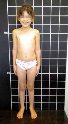
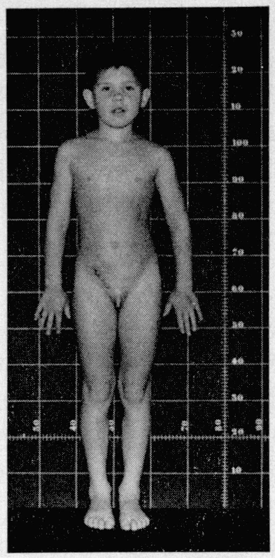
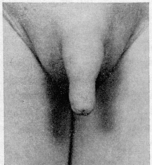
Patient with POR deficiency at 9 years old and her external genitalia appearance. 5 Imágenes
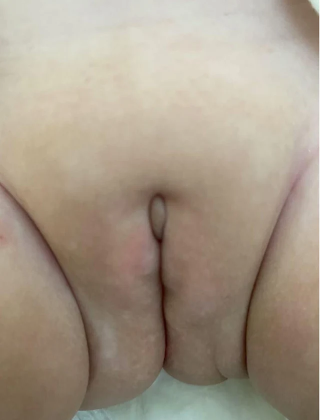
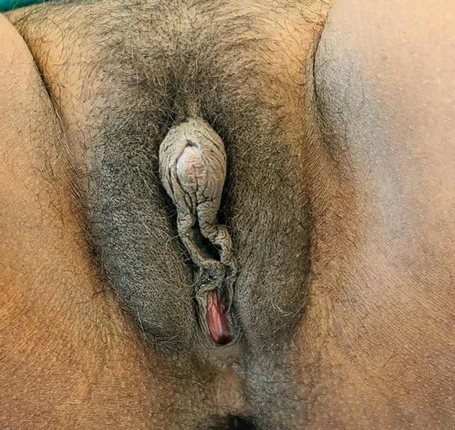
Full-body photograph of the 9-year-old girl with POR deficiency (stature grid background). External genitalia appearance of the POR deficiency patient. "Hammered silver" appearance associated with craniosynostosis (face at 9 years). Figure 1. Mother´s (A) and child’s (B) external genitalia 8 months after delivery. A B

DSD-C016

46,XX DSD; déficit de la citocromo P450 oxidorreductasa (POR) con síndrome de Antley-Bixler. · 46,XX · DSD-C016

46,XX · HPO:0000135, Hypogonadism (HP:0000135)

Por lo tanto, sobre la base de la virilización grave de la madre durante el embarazo, la primera hipótesis diagnóstica considerada fue el déficit de CYP19. Sin embargo, el resultado del análisis del gen CYP19 no confirmó ese diagnóstico.

CasoPublicación originalAtlas
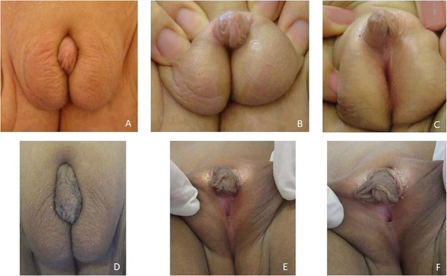
Fig. 2 The external genitalia of our patient at 10 months (A, B, C) and at 6 years old (D, E, F) 1 Imágenes

DSD-C017

46,XY DSD (déficit de 17β-hidroxiesteroide deshidrogenasa tipo 3, defecto de HSD17B3). · 46,XY · DSD-C017

AR, HSD17B3, NR5A1, SRD5A2 · 46,XY

46,XY DSD por defecto de HSD17B3.

CasoPublicación originalAtlas
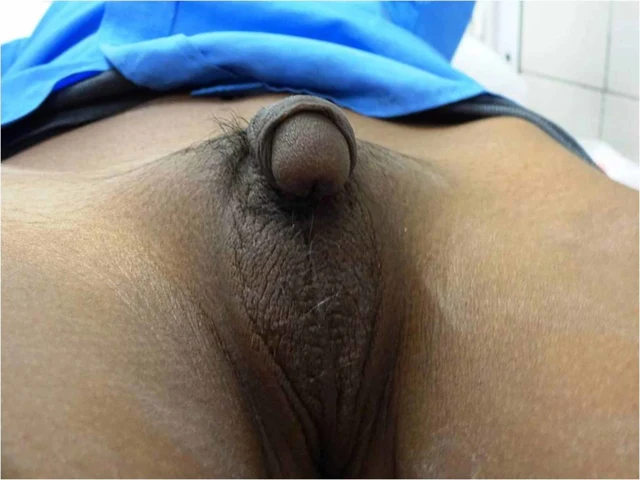
FIGURE 1: Clinical photograph showing fused labioscrotal folds with absent gonads, ventral chordee, and penoscrotal hypospadias Anthropometry revealed a weight of 17 kg, a height of 101 cm (more than 75th centile) with midparental height of 169 cm for a male child (between 25th and 50th centile) and 156 cm for a female child (less than 5th centile). Investigations revealed hypokalemia, primary adrenal insufficiency with elevated sex steroids, and elevated 11-deoxycortisol ( 2 Imágenes
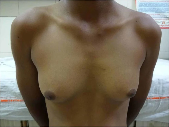
FIGURE 2: Clinical photograph showing Tanner stage 3 breast development There was no complaint of cyclical abdominal pain or hematuria. At this stage, the child was re-admitted and underwent definitive hysterectomy and bilateral salpingo-oophorectomy. This was followed by subcutaneous mastectomy and chordee correction. The child is now planned for a second stage surgery for hypospadias correction, following which testosterone replacement therapy will be initiated.

DSD-C018

46,XX DSD (hiperplasia suprarrenal congénita, déficit de 11β-hidroxilasa). · 46,XX · DSD-C018

46,XX · HPO:0000047, Hypospadias (HP:0000047)

El niño fue criado como varón por la familia y se le asignó un nombre masculino correspondiente. La exploración física mostró presión arterial en decúbito supino de 144–154/90–94 mmHg (por encima del percentil 99 para su edad y talla, compatible con el diagnóstico de hipertensión).

CasoPublicación originalAtlas
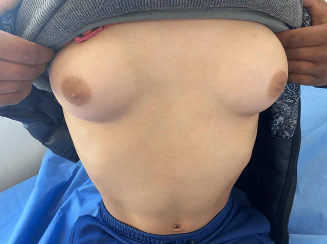
FIGURE 1: Clinical image showing breast development during central precocious puberty (Tanner stage 4). 2025 Hamaichat et al. Cureus 17(5): e83965. DOI 10.7759/cureus.83965 3 of 7 2 Imágenes
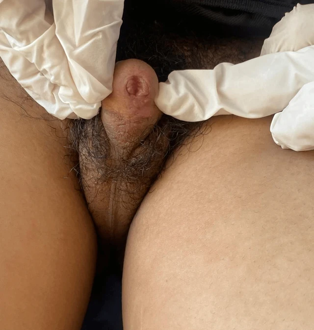
FIGURE 2: Clinical image showing ambiguous genitalia at Prader stage 5 and pubic hair at Tanner stage 5. Parameter Value Weight -1.5 SD Height -2 SD Pubic hair Tanner stage 5 Breast Tanner stage 4 External genitalia Prader stage 5 Follicle-stimulating hormone 4.48 mIU/mL (normal range: 2–13 mIU/mL) Luteinizing hormone 2.04 mIU/mL (normal range: 1.5–8 mIU/mL) Estradiol 52.23 pg/mL (34–186 pg/mL) Pelvic ultrasound Pubertal-sized uterus and a left lateral uterine structure 11 mm in height (left ovary), with the right ovary not visualized

DSD-C019

46,XX DSD (hiperplasia suprarrenal congénita, déficit de 11β-hidroxilasa). · 46,XX · DSD-C019

CYP11B1 · 46,XX · HPO:0000047, Hypospadias (HP:0000047)

Hiperplasia suprarrenal congénita (déficit de 11β-hidroxilasa); mujer 46,XX con virilización de genitales externos Prader 5 y pseudopubertad precoz.

CasoPublicación originalAtlas
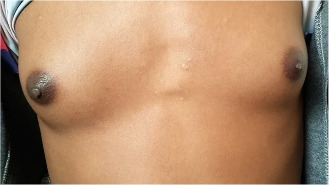
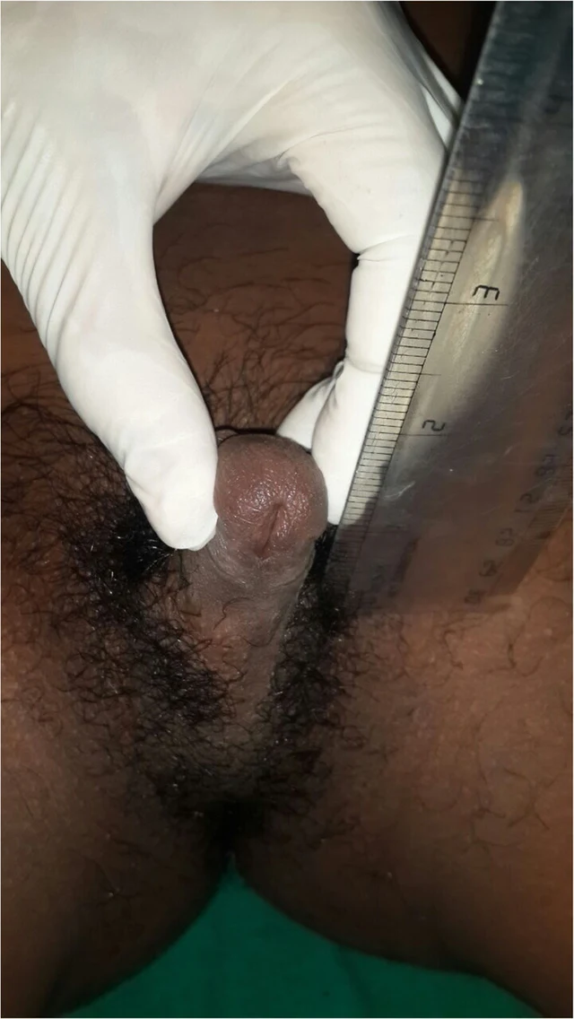
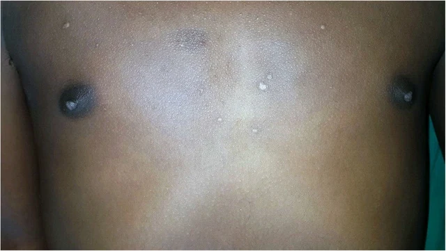
Imagen clínica · DSD-C02 3 Imágenes
Imagen clínica · DSD-C02 Imagen clínica · DSD-C02

DSD-C02

Hiperplasia suprarrenal congénita · 46,XX · DSD-C02

46,XX · HPO:0000047, HPO:0000771, Hypospadias (HP:0000047), Gynecomastia (HP:0000771)

Los autores establecieron el diagnóstico clínico de déficit de 11β-hidroxilasa según la hipertensión, la virilización y los resultados del eje suprarrenal; no se confirmó mediante genética ni 11-desoxicortisol.

CasoPublicación originalAtlas
Figure 5 Postoperative appearance of the external genitalia of twins. (A) Twin A at 30 months of age after feminizing surgery. (B) Twin B at 31 months of age after stage II hypospadias repair. associated with DSD, a group of rare conditions in which the chromosomal, gonadal, or phenotypical sex is incongruent (8). We herein present a case of 46,XY identical twins with DSD and UBMS caused by a novel heterozygous de novo splicing variant (c.1551-2A>G) in PPP1R12A. This variant results in the presence of a uterus in both 46,XY twins. Twin A exhibited gonadal dysgenesis and was assigned female, while Twin B had functional testes and was raised as male. PPP1R12A encodes PPP1R12A (1030aa), also named myosin phosphatase-targeting subunit 1 (MYPT1), expressed extensively and strongly in the brain, thyroid, 2 Imágenes
Figure 1 Appearance of the external genitalia of twins. (A,B) Twin A at 10 months of age. (C,D) Twin B at 30 months of age after stage I hypospadias repair. Testis-like masses were palpable in the area of stars.

DSD-C020

46,XY DSD · 46,XY · DSD-C020

46,XY

46,XY DSD asociado a variante de PPP1R12A con anomalías urogenitales e hipoplasia gonadal.

CasoPublicación originalAtlas
Figure 1 Appearance of the external genitalia of twins. (A,B) Twin A at 10 months of age. (C,D) Twin B at 30 months of age after stage I hypospadias repair. Testis-like masses were palpable in the area of stars. 2 Imágenes
Figure 5 Postoperative appearance of the external genitalia of twins. (A) Twin A at 30 months of age after feminizing surgery. (B) Twin B at 31 months of age after stage II hypospadias repair. associated with DSD, a group of rare conditions in which the chromosomal, gonadal, or phenotypical sex is incongruent (8). We herein present a case of 46,XY identical twins with DSD and UBMS caused by a novel heterozygous de novo splicing variant (c.1551-2A>G) in PPP1R12A. This variant results in the presence of a uterus in both 46,XY twins. Twin A exhibited gonadal dysgenesis and was assigned female, while Twin B had functional testes and was raised as male. PPP1R12A encodes PPP1R12A (1030aa), also named myosin phosphatase-targeting subunit 1 (MYPT1), expressed extensively and strongly in the brain, thyroid,

DSD-C021

DSD 46,XY (diferencias del desarrollo sexual) · 46,XY · DSD-C021

46,XY · HPO:0000047, Hypospadias (HP:0000047)

DSD 46,XY asociado a variante en PPP1R12A con anomalías del tracto urogenital.

CasoPublicación originalAtlas
FIGURE 4: Intraoperative appearance of bilateral gonadal structures. Surgical exploration demonstrating dysgenetic gonadal tissue located in the inguinal region. Histopathological examination revealed dysgenetic testicular tissue with a limited number of medium-sized seminiferous tubules showing maturation arrest at the primary spermatocyte stage, sparse Leydig cells, and absence of ovarian parenchyma or stroma. These findings were consistent with dysgenetic testicular tissue. Management and follow-up Management was discussed in a multidisciplinary setting involving pediatric endocrinologists, pediatric surgeons, geneticists, and psychologists. Age-appropriate information was provided, and shared decision- making was conducted with the patient and her parents. The patient expressed a stable female gender identity. Bilateral orchiectomy was performed in accordance with the declared gender, with psychological support. Histological analysis of the resected gonads showed complete testicular atrophy without intratubular germ cell neoplasia or malignant features. Estrogen replacement therapy with 17β-estradiol was subsequently initiated, along with management of hyperandrogenism and regular endocrinological follow-up. 1 Imágenes

DSD-C022

DSD 46,XY (diferencias del desarrollo sexual) · 46,XY · DSD-C022

NR5A1 · 46,XY · HPO:0000135, Hypogonadism (HP:0000135)

Los parámetros de crecimiento muestran una talla de 164 cm (+1.5 SD), un peso de 75 kg (>+2 SD) y un índice de masa corporal de 28.2 kg/m², compatible con sobrepeso según las curvas de referencia de crecimiento femeninas.

CasoPublicación originalAtlas
Figure 1 Genital appearance at 4 months showing coarse rugosity and visibly enlarged clitoris, and then at 2 years of age showing fine rugosity over labia and clitoral enlargement partially covered by labial fullness (patient K). 1 Imágenes

DSD-C023

Hiperplasia suprarrenal congénita · DSD-C023

A pesar de que la ambigüedad genital se detectó al nacer, el diagnóstico de CAH (hiperplasia suprarrenal congénita) no se estableció hasta los 9 meses de edad, momento en que se inició el tratamiento con esteroides. Sus dos hermanas (15 y 5 años) y un hermano (11 años) también fueron diagnosticados de CAH virilizante clásica.

CasoPublicación originalAtlas
Figure 2 Genital appearance at age 3.2 years and then at 4 years showing almost normal female genitalia (patient N). Congenital adrenal hyperplasia 557 1 Imágenes

DSD-C024

Hiperplasia suprarrenal congénita · DSD-C024

A pesar de que la ambigüedad genital se detectó al nacer, el diagnóstico de CAH (hiperplasia suprarrenal congénita) no se estableció hasta los 9 meses de edad, momento en que se inició el tratamiento con esteroides. Sus dos hermanas (15 y 5 años) y un hermano (11 años) también fueron diagnosticados de CAH virilizante clásica.

CasoPublicación originalAtlas
Figure 3 Genital appearance at initial presentation (2.5 years) and 1 year later (patient A). 558 B. Kulshreshtha et al. 1 Imágenes

DSD-C025

Hiperplasia suprarrenal congénita · DSD-C025

A pesar de que la ambigüedad genital se detectó al nacer, el diagnóstico de CAH (hiperplasia suprarrenal congénita) no se estableció hasta los 9 meses de edad, momento en que se inició el tratamiento con esteroides. Sus dos hermanas (15 y 5 años) y un hermano (11 años) también fueron diagnosticados de CAH virilizante clásica.

CasoPublicación originalAtlas
Figure 4. Appearance of external genitalia of the patient with features of partial virilization, including a small phallic structure, partially fused labioscrotal folds, and an external urethral meatus located in the perineal region, below the opening of the surgically created vagina. 1 Imágenes

DSD-C026

46,XY DSD (síndrome de insensibilidad parcial a los andrógenos combinado con hiperplasia suprarrenal congénita, déficit de P450 oxidorreductasa). · 46,XY · DSD-C026

46,XY

Presentamos el caso de una paciente que actualmente tiene 26 años, diagnosticada en la primera infancia como paciente DSD debido a la presentación ambigua de los órganos genitourinarios. Los datos de la entrevista sobre su ascendencia no revelaron ninguna patología de importancia para el diagnóstico.

CasoPublicación originalAtlas
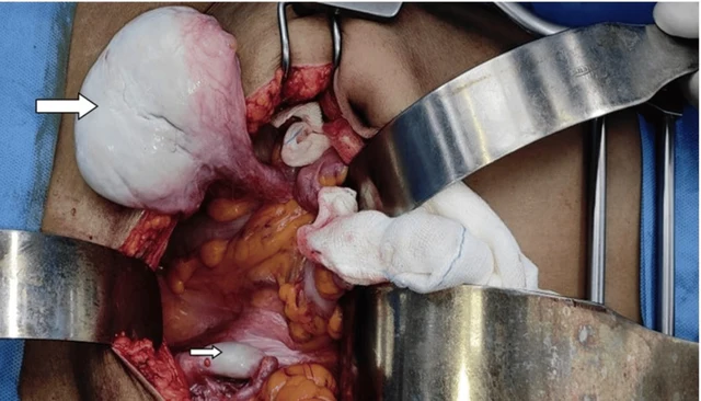
FIGURE 4: Testicular mass being separated from the local structures. Big arrow: Right testis, Small arrow: Left testis 2026 Ramya Lakshmi et al. Cureus 18(4): e106608. DOI 10.7759/cureus.106608 4 of 9 3 Imágenes
FIGURE 1: Absence of pubic hair growth FIGURE 2: Absence of axillary hair 2026 Ramya Lakshmi et al. Cureus 18(4): e106608. DOI 10.7759/cureus.106608 2 of 9

DSD-C027

Síndrome de insensibilidad a los andrógenos · 46,XY · DSD-C027

AR · 46,XY · HPO:0000786, Primary amenorrhea (HP:0000786)

Síndrome de insensibilidad completa a los andrógenos (CAIS); tumor germinal maligno originado en testículo intraabdominal (disgerminoma/seminoma, TxN3M0S1, estadio IIC); amenorrea primaria; ausencia de útero.

CasoPublicación originalAtlas
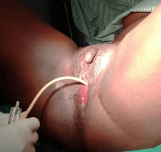
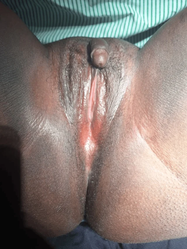
FIGURE 2: Case one catheterized At the six-week follow-up, the patient reported marked improvement in psychological well-being and satisfaction with the surgical outcome. Examination demonstrated a well-healed perineum and a functional vaginal canal measuring approximately 7 cm in length and 3 cm in width, as shown in Figure 3. She was also reviewed at three months and six months postoperatively, and she was happily sexually active without a complaint. Physical examination showed a long wide vaginal opening of about 7 cm long by 3 cm wide. 2026 Ainomugisha et al. Cureus 18(6): e111019. DOI 10.7759/cureus.111019 4 of 11 3 Imágenes
FIGURE 3: Case one six weeks post-surgery Case two A 25-year-old individual raised and socially recognized as male presented with progressive abdominal distension and lower abdominal pain of one year duration. During clinical evaluation, the patient disclosed the onset of virilization during adolescence, including the development of facial hair and a deepened voice, but also reported regular menstruation beginning at 15 years of age. The patient had been assigned male gender at birth because of the presence of a phallus, which had enlarged progressively with age. However, urine did not pass through the phallus. The patient identified socially as male and expressed a desire for relief from the abdominal swelling and cessation of menstruation. Both parents were deceased, and the patient was accompanied by a community health worker in whom he had previously confided. He had no formal education and survived on casual labor. On examination, the patient appeared chronically ill and wasted but demonstrated masculine body habitus, facial hair, a hoarse voice, smooth skin, and well-developed breasts. Vital signs were stable. Abdominal examination revealed gross distension with marked suprapubic tenderness. A large solid pelvic mass measuring approximately 18 × 10 cm was palpable, in addition to another irregular mass within the left lumbar region. Bilateral inguinal nodular swellings were also noted. Perineal examination demonstrated a phallus measuring approximately 6 × 3 cm without a urethral opening. A closed introitus with a small distal dimple was observed, through which urine was passed with straining in the lithotomy position. The stage, according to the Prader score, was 2. The anal opening was normally positioned. A provisional diagnosis of 2026 Ainomugisha et al. FIGURE 1: Case one before surgery A provisional diagnosis of ambiguous genitalia with suspected 46, XX DSD was made. Pelvic ultrasonography demonstrated a normal uterus, cervix, and right ovary. A cystic lesion measuring 33 × 4 × 20 mm with multiple internal echoes and a thick wall was identified within the left ovary. No abnormalities were detected in the urinary tract or upper abdominal organs. Due to financial limitations, karyotyping and hormonal assays could not be performed. The patient expressed a strong desire for surgical correction of the vaginal obstruction and removal of the enlarged clitoris after extensive counseling regarding anatomy, sexual function, and potential risks of surgery. Surgical Procedure Under spinal anesthesia and lithotomy positioning, examination under anesthesia confirmed a normal urethral opening located superior to the vaginal dimple. Catheterization, as shown in Figure 2, verified that the enlarged clitoris was not associated with the urinary tract. A diamond-shaped incision was created at the vaginal opening to enlarge the introitus. A vaginal canal approximately 6 cm long and 2 cm wide was identified, and the cervix was visualized. Vaginal mucosa was mobilized and anchored to the labial and perineal skin edges. A 20-mL syringe barrel was inserted and secured as a temporary vaginal stent. Clitoral reduction was subsequently performed by excision at the base near the mons pubis, followed by reconstruction and layered closure with meticulous hemostasis. Postoperatively, the patient remained hospitalized for 10 days and received antibiotics, analgesics, and perineal hygiene care. The urinary catheter 2026 Ainomugisha et al. Cureus 18(6): e111019. DOI 10.7759/cureus.111019 3 of 11

DSD-C028

46,XX DSD (diferencia del desarrollo sexual) · 46,XX · DSD-C028

46,XX

El autor informa genitales ambiguos, sospecha de 46,XX DSD.

CasoPublicación originalAtlas
Fig. 1. 7 years old girl with female pseudohermaphroditism due to virilizing adenoma of the mother. 2 Imágenes
Fig. 2. Male external genitalia of the same girl. The karyotype was 46XX. Serum electrolytes were within normal limits. The results of the steroid studies are presented below in more detail. A voiding cysto-urethrogram showed no anomalies. At surgery, the right inguinal canal was opened and no gonad was found. On revision of the abdominal cavity, two bean-sized gonads, two Fallopian tubes, and a small uterus attached to the bladder were detected. No Wolffian structures were found. A biopsy was taken from each gonad. Histological examination revealed ovarian tissue in both biopsies and 42 per cent of the

DSD-C029

46,XX DSD (pseudohermafroditismo femenino, exposición intrauterina a andrógenos por adenoma corticosuprarrenal materno). · 46,XX · DSD-C029

46,XX · HPO:0000028, Cryptorchidism (HP:0000028)

La paciente fue diagnosticada de pseudohermafroditismo femenino (46,XX, completamente masculinizada), causado por la secreción de andrógenos de un adenoma benigno de la corteza suprarrenal derecha materna; el diagnóstico anatomopatológico materno fue adenoma benigno de la corteza suprarrenal (65 g, 6,5×5×3 cm, sin células malignas).

CasoPublicación originalAtlas
Imagen clínica · DSD-C03 2 Imágenes
Imagen clínica · DSD-C03

DSD-C03

Déficit de aromatasa · 46,XX · DSD-C03

46,XX

Tras considerar los indicios de virilización durante el embarazo materno, el cariotipo 46,XX y la exclusión de las causas suprarrenales/testiculares habituales, los autores consideraron probable un déficit de aromatasa.

CasoPublicación originalAtlas
Fig. 4 Surgical management of ambiguous genitalia: third step, creation of a vagina 8 Imágenes
Fig. 1 a The preoperative aspect of ambiguous genitalia. b The design of further surgical incisions Fig. 3 Surgical management of ambiguous genitalia: second step, removing the erectile tissue from the clitoris Fig. 2 Surgical management of ambiguous genitalia: first step, clitoris degloving 550 Aesth Plast Surg (2014) 38:549–553 123 Fig. 6 Surgical management of ambiguous genitalia: fifth step, creation of a neovulva through tailoring of the labia minora using the dorsal clitoridean skin Aesth Plast Surg (2014) 38:549–553 551 123 Fig. 5 Surgical management of ambiguous genitalia: fourth step, retrourethral placement of the vagina Fig. 8 The aspect of ambiguous genitalia, postoperatively (a) and 2 years after surgery (b) 552 Aesth Plast Surg (2014) 38:549–553 123 Fig. 7 Surgical management of ambiguous genitalia: last step, fixing the flaps with separate sutures

DSD-C030

46,XX DSD (hiperplasia suprarrenal congénita, déficit de 21-hidroxilasa, forma clásica/perdedora de sal). · 46,XX · DSD-C030

46,XX

Antecedentes personales y medicación: una niña de 3 años acude por CAH clásica (debida a déficit de 21-hidroxilasa), diagnosticada a las 6 semanas de vida. El nivel elevado de 17-OHP sérica (150 nmol/L) apoya el diagnóstico de CAH (perdedora de sal).

CasoPublicación originalAtlas
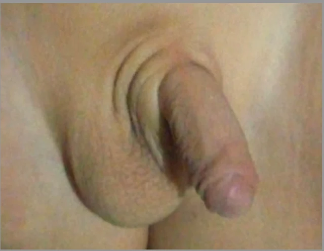
Figure 1: Patient’s abnormal genital phenotype: at the age of three days, (a) labia-majora-like bifid scrotum with clitoris-like microphallus and (b) perineoscrotal hypospadias; at the age of two years and three months, (c, d) microphallus had grown to 1.5 cm in length. 1 Imágenes

DSD-C031

Deficiencia de 5α-reductasa · 46,XY · DSD-C031

SRD5A2, SRY · 46,XY · HPO:0000047, HPO:0000054, Micropenis (HP:0000054), Hypospadias (HP:0000047)

46,XY DSD por deficiencia de 5α-reductasa-2; análisis molecular reveló variantes compuestas heterocigóticas de SRD5A2 c.485A>C (p.His162Pro) y c.680G>A (p.Arg227Gln).
CasoPublicación originalAtlas
Cargar más casos

Desliza a izquierda o derecha para ver las imágenes de este caso
Abrir imagen original
CasoPublicación originalAtlasVer el pie original en chino
DSD Research Library Versión de los datos v1.4.0