Aller au contenu
DSD Research Library Plateforme publique de recherche
Français
  • 简体中文
  • English
  • Français
  • Русский
  • Español
  • Português
Recherche Cas cliniques Recherche Atlas Publications Formation Méthodes

Galerie de cas

Explorez les cas et les figures de la collection originale. Recherchez par affection, gène, caryotype ou phénotype et consultez la publication source.

280 Cas illustrés571 Images

Dossiers de casGalerie de cas 280
Tout

Sélection actuelle: 280 Cas illustrés · 571 Images

Sélectionnez une image pour l’agrandir. Les images originales conservent le fichier source complet.

Image clinique · DSD-C01 3 Images
Image clinique · DSD-C01 Image clinique · DSD-C01

DSD-C01

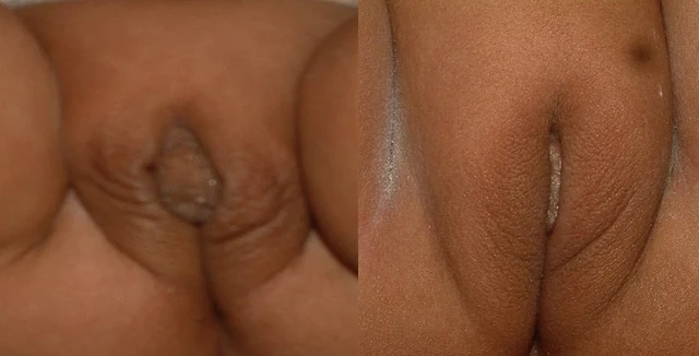
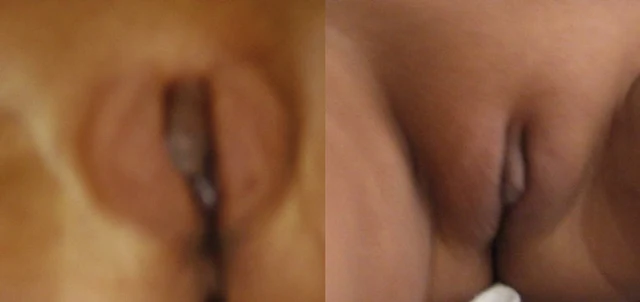
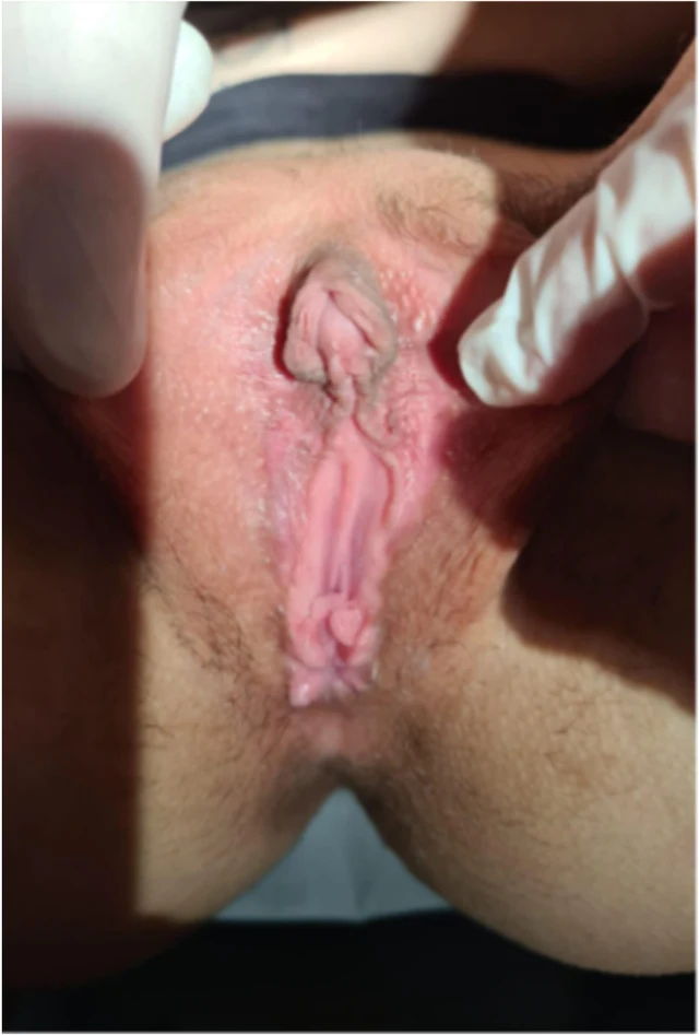
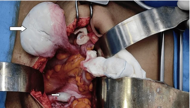
DSD ovotesticulaire · 46,XX · DSD-C01

46,XX · HPO:0000023, HPO:0000047, HPO:0000062, Inguinal hernia (HP:0000023), Ambiguous genitalia (HP:0000062), Hypospadias (HP:0000047)

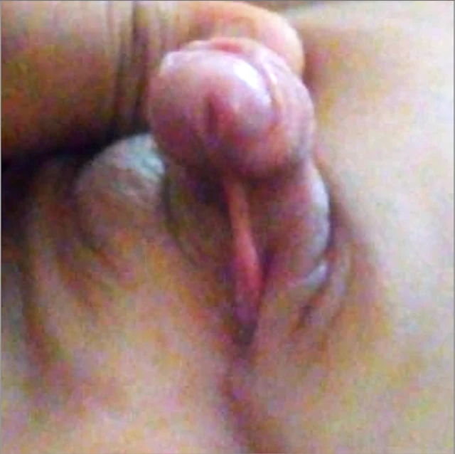
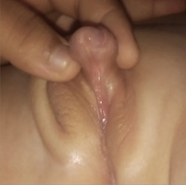
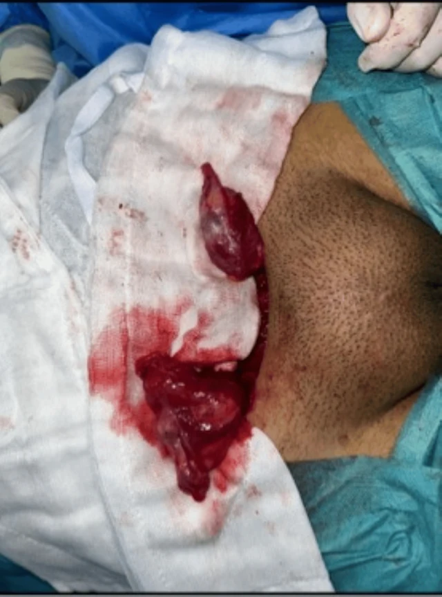
Ovotestis bipolaire droit, avec environ 50 % de tissu ovarien et 50 % de tissu testiculaire ; aucune tumeur à l'examen anatomopathologique ; score de potentiel de fertilité du tissu testiculaire de 0.

CasPublication sourceAtlas
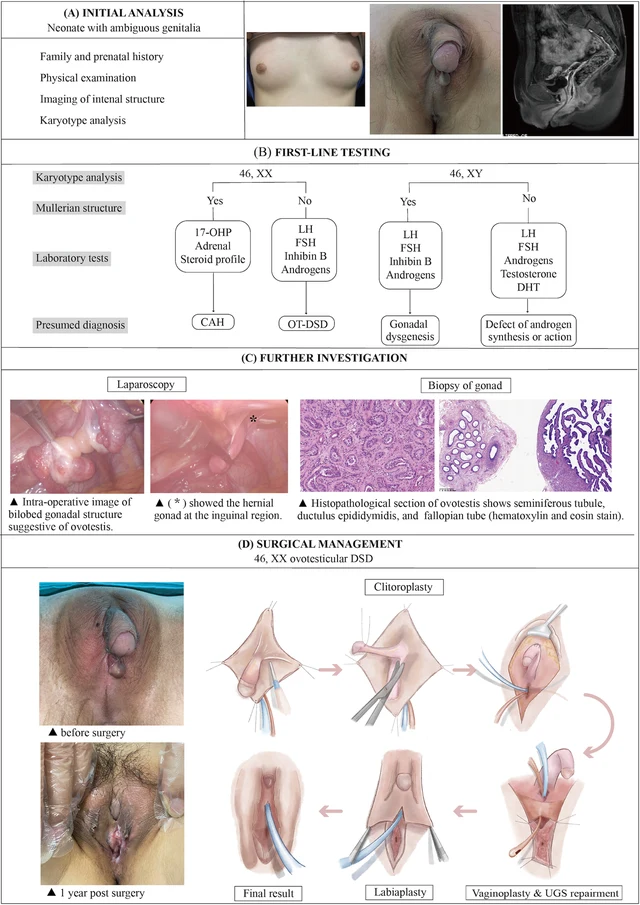
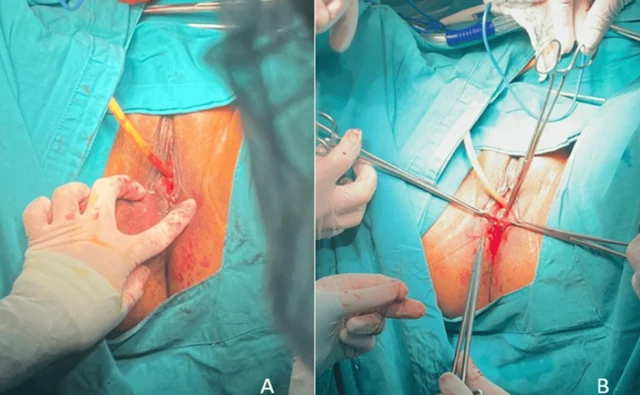
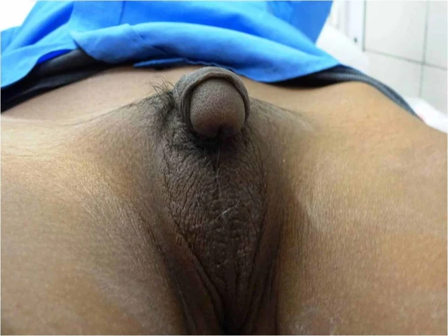
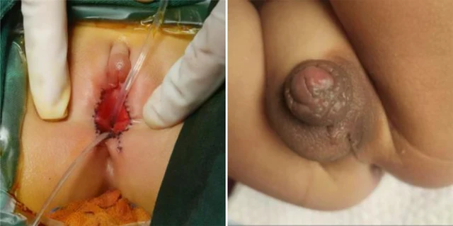
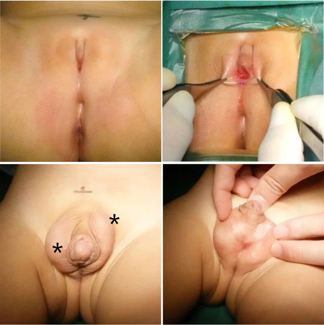
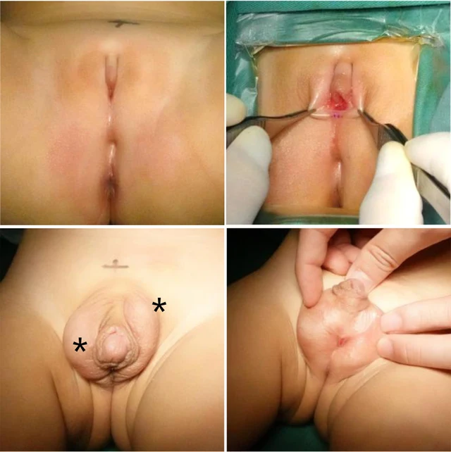
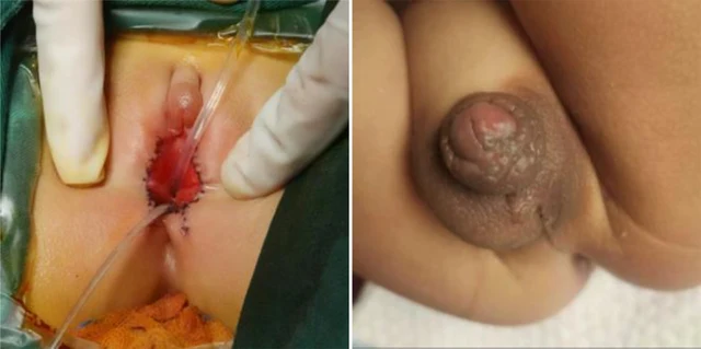
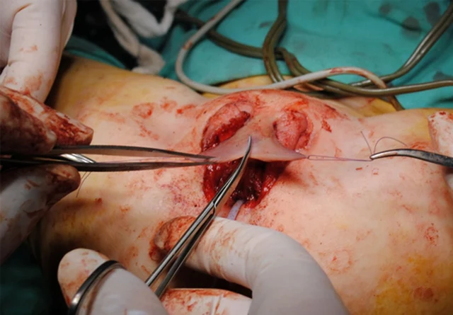
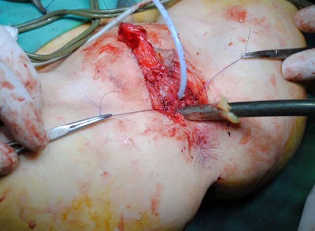
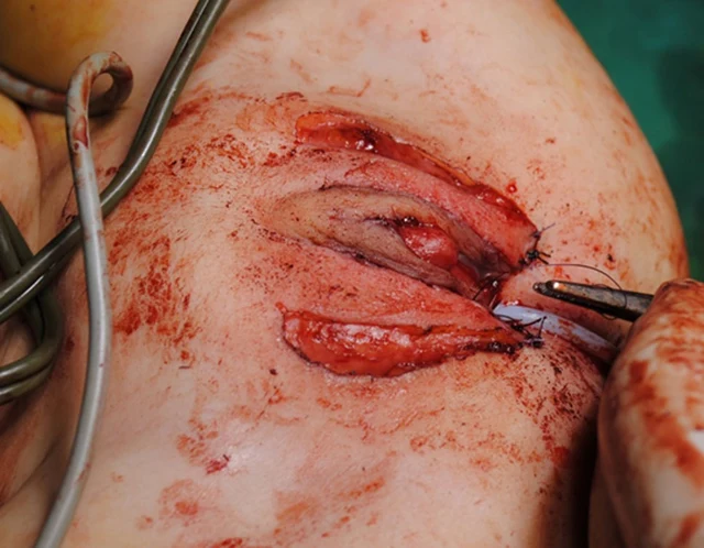
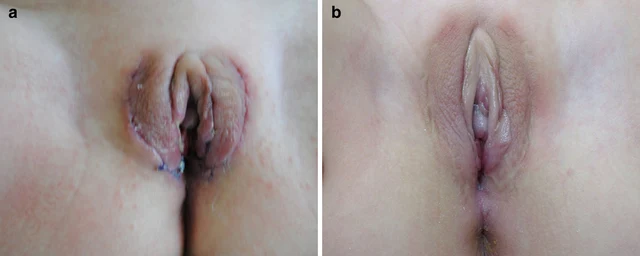
FIGURE 4 The illustration of the systematic diagnostic and treatment process for OT-DSD. The investigation process of OT-DSD can be summarized as initial assessment (A), first-line testing (B), and further investigation (C,D) showed the procedures of feminizing genitoplasty and pre- and 1-year post- operative pictures. The procedures included clitoroplasty, vaginoplasty and urogenital sinus repair, along with labiaplasty. 17, OHP, 17-alpha hydroxy progesterone; LH, Luteinizing hormone; FSH, follicle-stimulating hormone; DHT, dihydrotestosterone; CAH, congenital adrenal hyperplasia; OT-DSD, ovotesticular disorder of sex development. Zhao et al. 10.3389/fsurg.2024.1502340 Frontiers in Surgery 06 frontiersin.org 4 Images
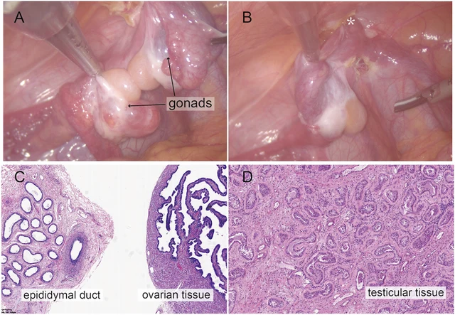
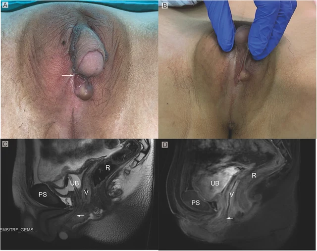
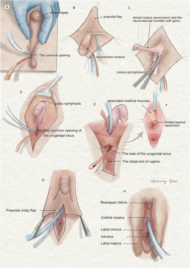
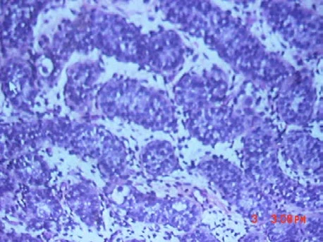
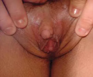
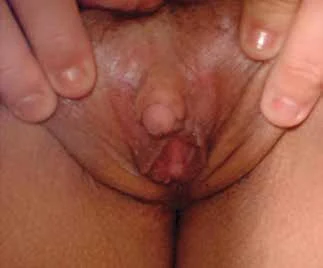
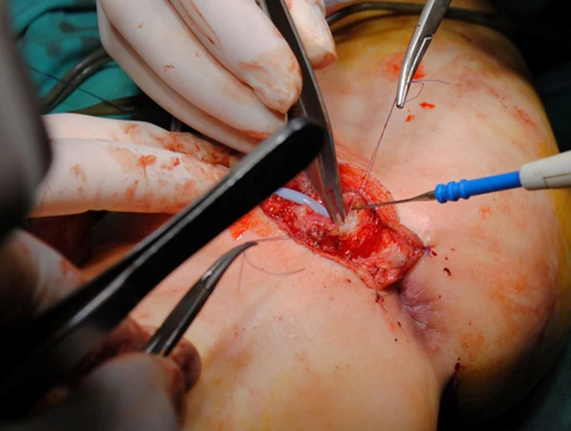
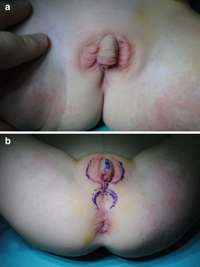
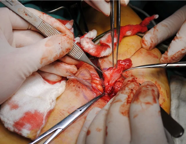
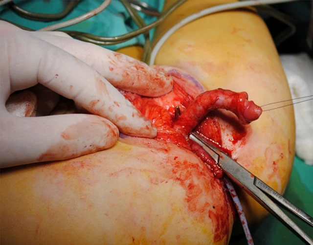
FIGURE 2 Intraoperative findings from laparoscopic exploration. (A) Intra-operative image of heterogenous gonadal structure showed the testicular and ovarian fractions organized in an end-to-end fashion, suggesting the ovotestis. (B) The right gonad was found at the inner ring of inguinal canal (*). (C) A biopsy was taken from the right hernial gonad in order to rule out any dysgenesis or ovotesticular tissue. Histopathological examination of sample 1 revealed epididymal and ovarian tissue. (D) Histopathological examination of sample 2 showed testicular histology with seminiferous tubules. Zhao et al. 10.3389/fsurg.2024.1502340 Frontiers in Surgery 03 frontiersin.org FIGURE 1 (A) Phenotypic appearance of genitalia before surgery. An enlarged clitoris, completely fused labia majora, and a movable painless mass at the right inguinal region was found. (B) An opening of the urogenital sinus was seen at the base of the clitoris. (C,D) Preoperative contrast-enhanced MRI showing well-defined urogenital sinus. Uterus was not detected. Sagittal MRI scan showing urinary bladder (UB), vagina (V), the common opening of the urogenital sinus (arrow), and rectum (R) The distance between the conference point to the superficial perineal pouch was below the level of the distal end of pubic symphysis (PS). Zhao et al. 10.3389/fsurg.2024.1502340 Frontiers in Surgery 02 frontiersin.org FIGURE 3 The illustration of the surgical procedures of feminizing genitoplasty. (A) The vulva showed Prader 4 virilization. (B) A subcoronal circumferential incision was made to de-glove the clitoris over Buck’s fascia. (C) The corporal bodies were excised with preservation of the glans clitoris and neurovascular bundles. (D) The corpora cavernosa was folded and sutured to the pubic arch. (E) The orange urethral catheter entered from the orifice through the urogenital sinus to exit from the fistula. (F) The fistula was sutured and secured with the rotating urethral mucosa flap. The vagina was pull-down to the vulve and sutured to the skin. (G) The prepuce in an onlay fashion covers the area between the clitoris and the vestibule. (H) The final result of the procedure. Zhao et al. 10.3389/fsurg.2024.1502340 Frontiers in Surgery 05 frontiersin.org

DSD-C011

DSD ovotesticulaire 46,XX (OT-DSD). · 46,XX · DSD-C011

46,XX · HPO:0000047, Hypospadias (HP:0000047)

Examens diagnostiques complémentaires : l'urétrocystoscopie a confirmé les constatations de l'IRM. Consultation multidisciplinaire : réunion multidisciplinaire (RCP) centrée sur le diagnostic et la décision chirurgicale, avec participation des services de chirurgie plastique, d'endocrinologie, de gynécologie, d'urologie, de gynécologie, de psychiatrie et du service social.

CasPublication sourceAtlas
FIGURE 1 Genitalia abnormalities (a) & (b) External genitalia at 3 weeks. (c) Genitocystography. (d) Testicular biopsy a b c d 1 Images

DSD-C012

DSD 46,XY (dysgénésie gonadique, délétion interstitielle 8p23.1). · 46,XY · DSD-C012

AR, GATA4, SRD5A2, WT1 · 46,XY · HPO:0000028, HPO:0000047, HPO:0000054, Micropenis (HP:0000054), Hypospadias (HP:0000047), Cryptorchidism (HP:0000028)

Diagnostic établi uniquement par caryotype à haute résolution : délétion interstitielle de la bande 8p23. Cette délétion est fréquente chez les patients présentant un retard psychomoteur et des malformations cardiaques, mais ne survient typiquement pas chez les patients atteints de malformations urogénitales primaires ; la littérature montre que ces malformations sont rapportées chez près d'un garçon sur deux.

CasPublication sourceAtlas
FIGURE 3: (A, B) CO2 Laser Vaginoplasty Psychological support was provided due to the potential impact of this condition on personal identity, perceptions of fertility, and sexual life. The psychological intervention helped the patient cope with the emotional and social challenges associated with the diagnosis, significantly improving her emotional well- being (Figure 4). 2024 Suárez Garcia et al. Cureus 16(11): e72895. DOI 10.7759/cureus.72895 5 of 9 1 Images

DSD-C013

DSD autre/non classé · 46,XX · DSD-C013

46,XX · HPO:0000135, Hypogonadism (HP:0000135)

Lors de la manœuvre de Valsalva, une imperforation de l'hymen a été constatée, confirmée par l'insertion d'un écouvillon et par la palpation. La patiente a été adressée en génétique afin d'explorer les diagnostics différentiels, notamment le mosaïcisme du syndrome de Turner et l'imperforation de l'hymen.

CasPublication sourceAtlas
Figure 1 The characteristics of the external genitalia of the proband at the age of 5 months. Ambiguous external genitalia was observed in the proband, with a 0.5 cm penis, a urethral opening in the perineum, a split scrotum resembling labia majora, and testicles found in the inguinal region. 1 Images

DSD-C014

DSD 46,XY · 46,XY · DSD-C014

NR0B1 · 46,XY

DSD 46,XY associé à une duplication Xp21 et dystrophie musculaire.

CasPublication sourceAtlas
Figure 1 Clitoromegaly of 3 cm length and 1 cm breadth with clitoral index of 300 m2. 2 JCEM Case Reports, 2026, Volume 4, Issue 7 1 Images

DSD-C015

Hyperplasie congénitale des surrénales · 46,XX · DSD-C015

CYP21A2 · 46,XX · HPO:0004322, Short stature (HP:0004322)

Après le diagnostic initial, ses parents ne sont plus revenus en consultation ni n'ont demandé un second avis pour l'aménorrhée. Évaluation diagnostique : l'examen physique montre une petite taille (140,5 cm ; taille médiane des parents 151 cm, −3,1 SDS), un poids de 42 kg, un indice de masse corporelle de 21 kg/m², une pression artérielle et des proportions corporelles normales, un stade de Tanner A4B3P4.

CasPublication sourceAtlas
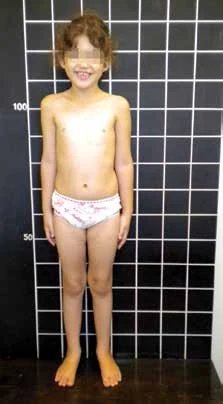
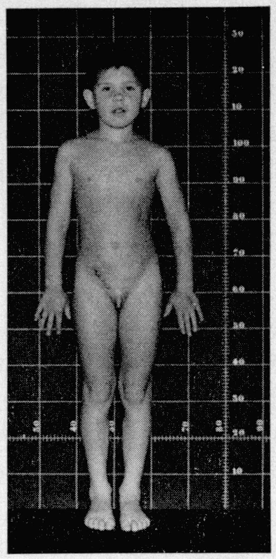
Patient with POR deficiency at 9 years old and her external genitalia appearance. 5 Images
Full-body photograph of the 9-year-old girl with POR deficiency (stature grid background). External genitalia appearance of the POR deficiency patient. "Hammered silver" appearance associated with craniosynostosis (face at 9 years). Figure 1. Mother´s (A) and child’s (B) external genitalia 8 months after delivery. A B

DSD-C016

DSD 46,XX ; déficit en cytochrome P450 oxydoréductase (POR) associé au syndrome d'Antley-Bixler. · 46,XX · DSD-C016

46,XX · HPO:0000135, Hypogonadism (HP:0000135)

Par conséquent, en raison de la virilisation sévère de la mère pendant la grossesse, l'hypothèse diagnostique principale envisagée était le déficit en CYP19. Toutefois, les résultats de l'analyse du gène CYP19 n'ont pas confirmé ce diagnostic.

CasPublication sourceAtlas
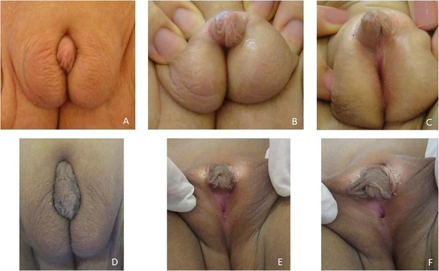
Fig. 2 The external genitalia of our patient at 10 months (A, B, C) and at 6 years old (D, E, F) 1 Images

DSD-C017

DSD 46,XY (déficit en 17β-hydroxystéroïde déshydrogénase de type 3, déficit en HSD17B3). · 46,XY · DSD-C017

AR, HSD17B3, NR5A1, SRD5A2 · 46,XY

DSD 46,XY dû à un déficit en HSD17B3.

CasPublication sourceAtlas
FIGURE 1: Clinical photograph showing fused labioscrotal folds with absent gonads, ventral chordee, and penoscrotal hypospadias Anthropometry revealed a weight of 17 kg, a height of 101 cm (more than 75th centile) with midparental height of 169 cm for a male child (between 25th and 50th centile) and 156 cm for a female child (less than 5th centile). Investigations revealed hypokalemia, primary adrenal insufficiency with elevated sex steroids, and elevated 11-deoxycortisol ( 2 Images
FIGURE 2: Clinical photograph showing Tanner stage 3 breast development There was no complaint of cyclical abdominal pain or hematuria. At this stage, the child was re-admitted and underwent definitive hysterectomy and bilateral salpingo-oophorectomy. This was followed by subcutaneous mastectomy and chordee correction. The child is now planned for a second stage surgery for hypospadias correction, following which testosterone replacement therapy will be initiated.

DSD-C018

DSD 46,XX (hyperplasie congénitale des surrénales, déficit en 11β-hydroxylase). · 46,XX · DSD-C018

46,XX · HPO:0000047, Hypospadias (HP:0000047)

L'enfant a été élevé comme un garçon par la famille et a reçu un prénom masculin concordant. À l'examen, la pression artérielle en décubitus dorsal était de 144–154/90–94 mmHg (supérieure au 99e centile pour l'âge et la taille, compatible avec le diagnostic d'hypertension).

CasPublication sourceAtlas
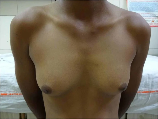
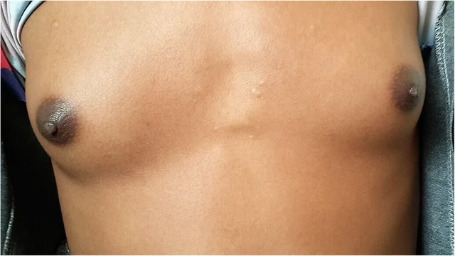
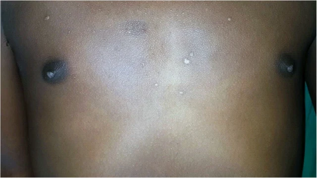
FIGURE 1: Clinical image showing breast development during central precocious puberty (Tanner stage 4). 2025 Hamaichat et al. Cureus 17(5): e83965. DOI 10.7759/cureus.83965 3 of 7 2 Images
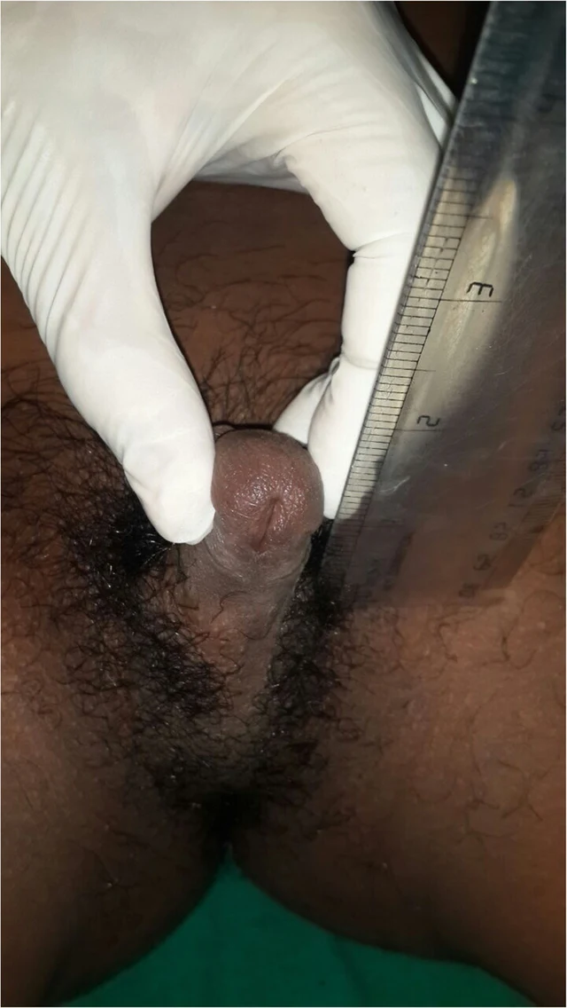
FIGURE 2: Clinical image showing ambiguous genitalia at Prader stage 5 and pubic hair at Tanner stage 5. Parameter Value Weight -1.5 SD Height -2 SD Pubic hair Tanner stage 5 Breast Tanner stage 4 External genitalia Prader stage 5 Follicle-stimulating hormone 4.48 mIU/mL (normal range: 2–13 mIU/mL) Luteinizing hormone 2.04 mIU/mL (normal range: 1.5–8 mIU/mL) Estradiol 52.23 pg/mL (34–186 pg/mL) Pelvic ultrasound Pubertal-sized uterus and a left lateral uterine structure 11 mm in height (left ovary), with the right ovary not visualized

DSD-C019

DSD 46,XX (hyperplasie congénitale des surrénales, déficit en 11β-hydroxylase). · 46,XX · DSD-C019

CYP11B1 · 46,XX · HPO:0000047, Hypospadias (HP:0000047)

Hyperplasie congénitale des surrénales (déficit en 11β-hydroxylase) ; sujet féminin 46,XX, virilisation des organes génitaux externes de stade 5 de Prader, pseudo-puberté précoce.

CasPublication sourceAtlas
Image clinique · DSD-C02 3 Images
Image clinique · DSD-C02 Image clinique · DSD-C02

DSD-C02

Hyperplasie congénitale des surrénales · 46,XX · DSD-C02

46,XX · HPO:0000047, HPO:0000771, Hypospadias (HP:0000047), Gynecomastia (HP:0000771)

Les auteurs ont posé un diagnostic clinique de déficit en 11β-hydroxylase sur la base de l'hypertension, de la virilisation et des résultats de l'axe surrénalien ; aucune confirmation génétique ni par le 11-désoxycortisol n'a été réalisée.

CasPublication sourceAtlas
Figure 5 Postoperative appearance of the external genitalia of twins. (A) Twin A at 30 months of age after feminizing surgery. (B) Twin B at 31 months of age after stage II hypospadias repair. associated with DSD, a group of rare conditions in which the chromosomal, gonadal, or phenotypical sex is incongruent (8). We herein present a case of 46,XY identical twins with DSD and UBMS caused by a novel heterozygous de novo splicing variant (c.1551-2A>G) in PPP1R12A. This variant results in the presence of a uterus in both 46,XY twins. Twin A exhibited gonadal dysgenesis and was assigned female, while Twin B had functional testes and was raised as male. PPP1R12A encodes PPP1R12A (1030aa), also named myosin phosphatase-targeting subunit 1 (MYPT1), expressed extensively and strongly in the brain, thyroid, 2 Images
Figure 1 Appearance of the external genitalia of twins. (A,B) Twin A at 10 months of age. (C,D) Twin B at 30 months of age after stage I hypospadias repair. Testis-like masses were palpable in the area of stars.

DSD-C020

DSD 46,XY · 46,XY · DSD-C020

46,XY

DSD 46,XY associé à un variant de PPP1R12A avec anomalies urogénitales et dysgénésie gonadique.

CasPublication sourceAtlas
Figure 1 Appearance of the external genitalia of twins. (A,B) Twin A at 10 months of age. (C,D) Twin B at 30 months of age after stage I hypospadias repair. Testis-like masses were palpable in the area of stars. 2 Images
Figure 5 Postoperative appearance of the external genitalia of twins. (A) Twin A at 30 months of age after feminizing surgery. (B) Twin B at 31 months of age after stage II hypospadias repair. associated with DSD, a group of rare conditions in which the chromosomal, gonadal, or phenotypical sex is incongruent (8). We herein present a case of 46,XY identical twins with DSD and UBMS caused by a novel heterozygous de novo splicing variant (c.1551-2A>G) in PPP1R12A. This variant results in the presence of a uterus in both 46,XY twins. Twin A exhibited gonadal dysgenesis and was assigned female, while Twin B had functional testes and was raised as male. PPP1R12A encodes PPP1R12A (1030aa), also named myosin phosphatase-targeting subunit 1 (MYPT1), expressed extensively and strongly in the brain, thyroid,

DSD-C021

DSD 46,XY (différences du développement sexuel) · 46,XY · DSD-C021

46,XY · HPO:0000047, Hypospadias (HP:0000047)

DSD 46,XY associé à une variante de PPP1R12A avec anomalies urogénitales.

CasPublication sourceAtlas
FIGURE 4: Intraoperative appearance of bilateral gonadal structures. Surgical exploration demonstrating dysgenetic gonadal tissue located in the inguinal region. Histopathological examination revealed dysgenetic testicular tissue with a limited number of medium-sized seminiferous tubules showing maturation arrest at the primary spermatocyte stage, sparse Leydig cells, and absence of ovarian parenchyma or stroma. These findings were consistent with dysgenetic testicular tissue. Management and follow-up Management was discussed in a multidisciplinary setting involving pediatric endocrinologists, pediatric surgeons, geneticists, and psychologists. Age-appropriate information was provided, and shared decision- making was conducted with the patient and her parents. The patient expressed a stable female gender identity. Bilateral orchiectomy was performed in accordance with the declared gender, with psychological support. Histological analysis of the resected gonads showed complete testicular atrophy without intratubular germ cell neoplasia or malignant features. Estrogen replacement therapy with 17β-estradiol was subsequently initiated, along with management of hyperandrogenism and regular endocrinological follow-up. 1 Images

DSD-C022

DSD 46,XY (différences du développement sexuel) · 46,XY · DSD-C022

NR5A1 · 46,XY · HPO:0000135, Hypogonadism (HP:0000135)

Les paramètres de croissance montrent une taille de 164 cm (+1.5 SD), un poids de 75 kg (>+2 SD) et un indice de masse corporelle de 28.2 kg/m², compatibles avec un surpoids selon les courbes de croissance de référence féminines.

CasPublication sourceAtlas
Figure 1 Genital appearance at 4 months showing coarse rugosity and visibly enlarged clitoris, and then at 2 years of age showing fine rugosity over labia and clitoral enlargement partially covered by labial fullness (patient K). 1 Images

DSD-C023

Hyperplasie congénitale des surrénales · DSD-C023

Ambiguïté génitale constatée à la naissance ; diagnostic de CAH posé à 9 mois avec début du traitement hormonal. Ses deux sœurs (15 ans et 5 ans) et son frère (11 ans) ont également reçu un diagnostic de CAH classique virilisante.

CasPublication sourceAtlas
Figure 2 Genital appearance at age 3.2 years and then at 4 years showing almost normal female genitalia (patient N). Congenital adrenal hyperplasia 557 1 Images

DSD-C024

Hyperplasie congénitale des surrénales · DSD-C024

Ambiguïté génitale constatée à la naissance ; diagnostic de CAH posé à 9 mois avec début du traitement hormonal. Ses deux sœurs (15 ans et 5 ans) et son frère (11 ans) ont également reçu un diagnostic de CAH classique virilisante.

CasPublication sourceAtlas
Figure 3 Genital appearance at initial presentation (2.5 years) and 1 year later (patient A). 558 B. Kulshreshtha et al. 1 Images

DSD-C025

Hyperplasie congénitale des surrénales · DSD-C025

Ambiguïté génitale constatée à la naissance ; diagnostic de CAH posé à 9 mois avec début du traitement hormonal. Ses deux sœurs (15 ans et 5 ans) et son frère (11 ans) ont également reçu un diagnostic de CAH classique virilisante.

CasPublication sourceAtlas
Figure 4. Appearance of external genitalia of the patient with features of partial virilization, including a small phallic structure, partially fused labioscrotal folds, and an external urethral meatus located in the perineal region, below the opening of the surgically created vagina. 1 Images

DSD-C026

DSD 46,XY (syndrome d'insensibilité partielle aux androgènes associé à une hyperplasie congénitale des surrénales, déficit en P450 oxydoréductase). · 46,XY · DSD-C026

46,XY

Nous rapportons le cas d'une patiente aujourd'hui âgée de 26 ans, diagnostiquée DSD au début de la petite enfance en raison d'une présentation ambiguë des organes génitourinaires. L'entretien sur son ascendance n'a révélé aucune pathologie importante pour le diagnostic.

CasPublication sourceAtlas
FIGURE 4: Testicular mass being separated from the local structures. Big arrow: Right testis, Small arrow: Left testis 2026 Ramya Lakshmi et al. Cureus 18(4): e106608. DOI 10.7759/cureus.106608 4 of 9 3 Images
FIGURE 1: Absence of pubic hair growth FIGURE 2: Absence of axillary hair 2026 Ramya Lakshmi et al. Cureus 18(4): e106608. DOI 10.7759/cureus.106608 2 of 9

DSD-C027

Syndrome d'insensibilité aux androgènes · 46,XY · DSD-C027

AR · 46,XY · HPO:0000786, Primary amenorrhea (HP:0000786)

Syndrome d'insensibilité complète aux androgènes (CAIS) ; tumeur germinale maligne d'origine testiculaire intrapéritonéale (dysgerminome/séminome, TxN3M0S1, stade IIC) ; aménorrhée primaire ; absence d'utérus.

CasPublication sourceAtlas
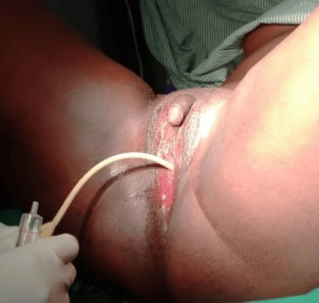
FIGURE 2: Case one catheterized At the six-week follow-up, the patient reported marked improvement in psychological well-being and satisfaction with the surgical outcome. Examination demonstrated a well-healed perineum and a functional vaginal canal measuring approximately 7 cm in length and 3 cm in width, as shown in Figure 3. She was also reviewed at three months and six months postoperatively, and she was happily sexually active without a complaint. Physical examination showed a long wide vaginal opening of about 7 cm long by 3 cm wide. 2026 Ainomugisha et al. Cureus 18(6): e111019. DOI 10.7759/cureus.111019 4 of 11 3 Images
FIGURE 3: Case one six weeks post-surgery Case two A 25-year-old individual raised and socially recognized as male presented with progressive abdominal distension and lower abdominal pain of one year duration. During clinical evaluation, the patient disclosed the onset of virilization during adolescence, including the development of facial hair and a deepened voice, but also reported regular menstruation beginning at 15 years of age. The patient had been assigned male gender at birth because of the presence of a phallus, which had enlarged progressively with age. However, urine did not pass through the phallus. The patient identified socially as male and expressed a desire for relief from the abdominal swelling and cessation of menstruation. Both parents were deceased, and the patient was accompanied by a community health worker in whom he had previously confided. He had no formal education and survived on casual labor. On examination, the patient appeared chronically ill and wasted but demonstrated masculine body habitus, facial hair, a hoarse voice, smooth skin, and well-developed breasts. Vital signs were stable. Abdominal examination revealed gross distension with marked suprapubic tenderness. A large solid pelvic mass measuring approximately 18 × 10 cm was palpable, in addition to another irregular mass within the left lumbar region. Bilateral inguinal nodular swellings were also noted. Perineal examination demonstrated a phallus measuring approximately 6 × 3 cm without a urethral opening. A closed introitus with a small distal dimple was observed, through which urine was passed with straining in the lithotomy position. The stage, according to the Prader score, was 2. The anal opening was normally positioned. A provisional diagnosis of 2026 Ainomugisha et al. FIGURE 1: Case one before surgery A provisional diagnosis of ambiguous genitalia with suspected 46, XX DSD was made. Pelvic ultrasonography demonstrated a normal uterus, cervix, and right ovary. A cystic lesion measuring 33 × 4 × 20 mm with multiple internal echoes and a thick wall was identified within the left ovary. No abnormalities were detected in the urinary tract or upper abdominal organs. Due to financial limitations, karyotyping and hormonal assays could not be performed. The patient expressed a strong desire for surgical correction of the vaginal obstruction and removal of the enlarged clitoris after extensive counseling regarding anatomy, sexual function, and potential risks of surgery. Surgical Procedure Under spinal anesthesia and lithotomy positioning, examination under anesthesia confirmed a normal urethral opening located superior to the vaginal dimple. Catheterization, as shown in Figure 2, verified that the enlarged clitoris was not associated with the urinary tract. A diamond-shaped incision was created at the vaginal opening to enlarge the introitus. A vaginal canal approximately 6 cm long and 2 cm wide was identified, and the cervix was visualized. Vaginal mucosa was mobilized and anchored to the labial and perineal skin edges. A 20-mL syringe barrel was inserted and secured as a temporary vaginal stent. Clitoral reduction was subsequently performed by excision at the base near the mons pubis, followed by reconstruction and layered closure with meticulous hemostasis. Postoperatively, the patient remained hospitalized for 10 days and received antibiotics, analgesics, and perineal hygiene care. The urinary catheter 2026 Ainomugisha et al. Cureus 18(6): e111019. DOI 10.7759/cureus.111019 3 of 11

DSD-C028

DSD 46,XX (différence du développement sexuel) · 46,XX · DSD-C028

46,XX

Les auteurs rapportent des organes génitaux ambigus, suspicion de DSD 46,XX.

CasPublication sourceAtlas
Fig. 1. 7 years old girl with female pseudohermaphroditism due to virilizing adenoma of the mother. 2 Images
Fig. 2. Male external genitalia of the same girl. The karyotype was 46XX. Serum electrolytes were within normal limits. The results of the steroid studies are presented below in more detail. A voiding cysto-urethrogram showed no anomalies. At surgery, the right inguinal canal was opened and no gonad was found. On revision of the abdominal cavity, two bean-sized gonads, two Fallopian tubes, and a small uterus attached to the bladder were detected. No Wolffian structures were found. A biopsy was taken from each gonad. Histological examination revealed ovarian tissue in both biopsies and 42 per cent of the

DSD-C029

DSD 46,XX (pseudohermaphrodisme féminin, exposition androgénique in utero due à un adénome corticosurrénalien maternel). · 46,XX · DSD-C029

46,XX · HPO:0000028, Cryptorchidism (HP:0000028)

La patiente a reçu le diagnostic de pseudohermaphrodisme féminin (46,XX, complètement virilisé), dû à la sécrétion d'androgènes par un adénome corticosurrénalien bénin droit maternel ; le diagnostic pathologique de la mère était un adénome corticosurrénalien bénin (65 g, 6,5 × 5 × 3 cm, sans cellules malignes).

CasPublication sourceAtlas
Image clinique · DSD-C03 2 Images
Image clinique · DSD-C03

DSD-C03

Déficit en aromatase · 46,XX · DSD-C03

46,XX

Compte tenu des signes de virilisation pendant la grossesse maternelle, du caryotype 46,XX et après exclusion des causes surrénaliennes/testiculaires courantes, les auteurs considèrent le déficit en aromatase comme probable.

CasPublication sourceAtlas
Fig. 4 Surgical management of ambiguous genitalia: third step, creation of a vagina 8 Images
Fig. 1 a The preoperative aspect of ambiguous genitalia. b The design of further surgical incisions Fig. 3 Surgical management of ambiguous genitalia: second step, removing the erectile tissue from the clitoris Fig. 2 Surgical management of ambiguous genitalia: first step, clitoris degloving 550 Aesth Plast Surg (2014) 38:549–553 123 Fig. 6 Surgical management of ambiguous genitalia: fifth step, creation of a neovulva through tailoring of the labia minora using the dorsal clitoridean skin Aesth Plast Surg (2014) 38:549–553 551 123 Fig. 5 Surgical management of ambiguous genitalia: fourth step, retrourethral placement of the vagina Fig. 8 The aspect of ambiguous genitalia, postoperatively (a) and 2 years after surgery (b) 552 Aesth Plast Surg (2014) 38:549–553 123 Fig. 7 Surgical management of ambiguous genitalia: last step, fixing the flaps with separate sutures

DSD-C030

DSD 46,XX (hyperplasie congénitale des surrénales, déficit en 21-hydroxylase, forme classique avec perte de sel). · 46,XX · DSD-C030

46,XX

Antécédents et traitement : une fillette de 3 ans consulte pour une HCS classique (due à un déficit en 21-hydroxylase), diagnostiquée à l'âge de 6 semaines. Une élévation du taux sérique de 17-OHP (150 nmol/L) soutient le diagnostic d'HCS (forme avec perte de sel).

CasPublication sourceAtlas
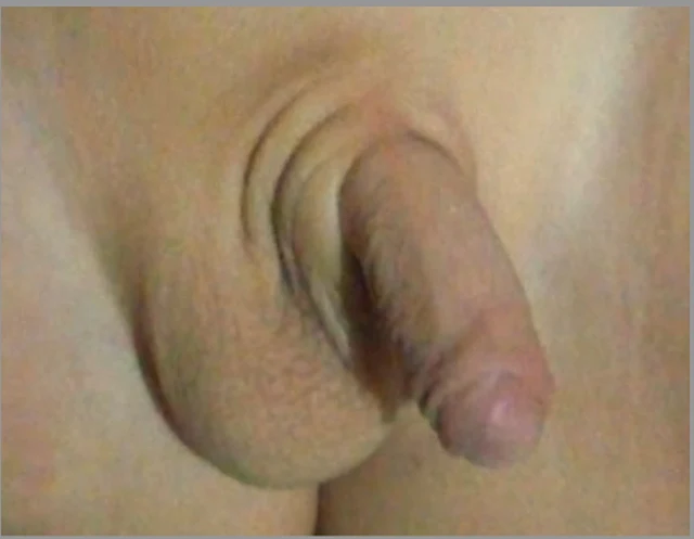
Figure 1: Patient’s abnormal genital phenotype: at the age of three days, (a) labia-majora-like bifid scrotum with clitoris-like microphallus and (b) perineoscrotal hypospadias; at the age of two years and three months, (c, d) microphallus had grown to 1.5 cm in length. 1 Images

DSD-C031

Déficit en 5α-réductase · 46,XY · DSD-C031

SRD5A2, SRY · 46,XY · HPO:0000047, HPO:0000054, Micropenis (HP:0000054), Hypospadias (HP:0000047)

DSD 46,XY dû à un déficit en 5α-réductase-2 ; l'analyse moléculaire a identifié des variants hétérozygotes composites du gène SRD5A2 : c.485A>C (p.His162Pro) et c.680G>A (p.Arg227Gln).
CasPublication sourceAtlas
Charger d’autres cas

Balayez à gauche ou à droite pour voir les images de ce cas
Ouvrir l’image originale
CasPublication sourceAtlasVoir la légende originale en chinois
DSD Research Library Version des données v1.4.0