Перейти к содержанию
DSD Research Library Открытая исследовательская платформа
Русский
  • 简体中文
  • English
  • Français
  • Русский
  • Español
  • Português
Поиск Регистр случаев Исследования Атлас Публикации Обучение Методы

Галерея случаев

Изучайте случаи и иллюстрации исходной коллекции. Ищите по заболеванию, гену, кариотипу или фенотипу и переходите к первоисточнику.

280 Случаи с изображениями571 Изображения

Список случаевГалерея случаев 280
Все

Текущая выборка: 280 Случаи с изображениями · 571 Изображения

Выберите изображение для увеличения. Исходные изображения сохраняют файл целиком.

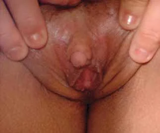
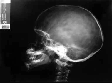
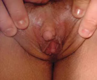
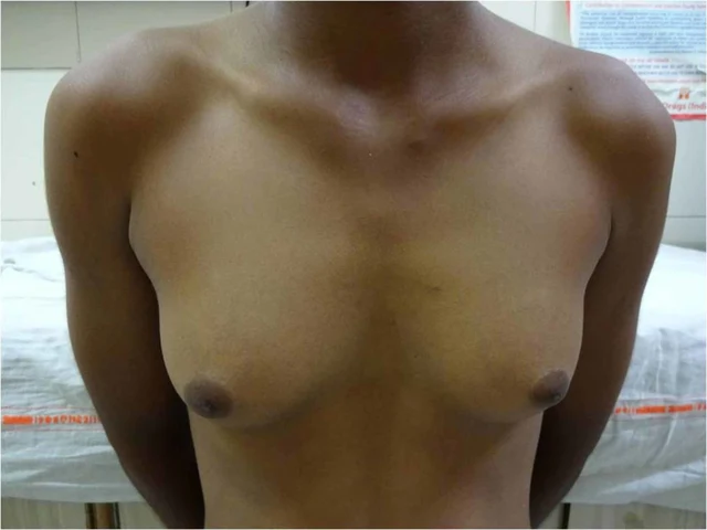
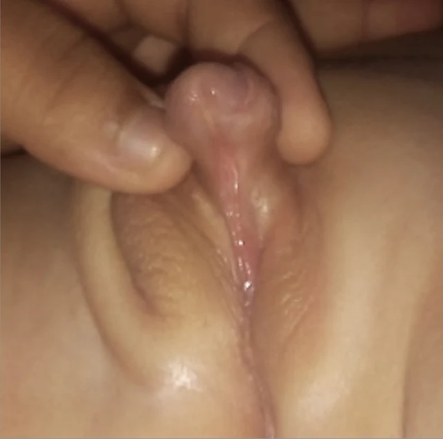
Клиническое изображение · DSD-C01 3 Изображения
Клиническое изображение · DSD-C01 Клиническое изображение · DSD-C01

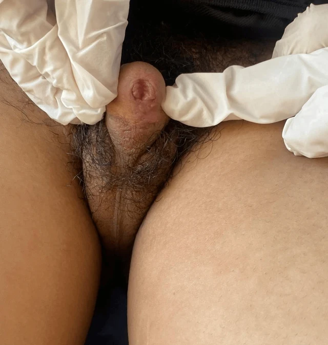
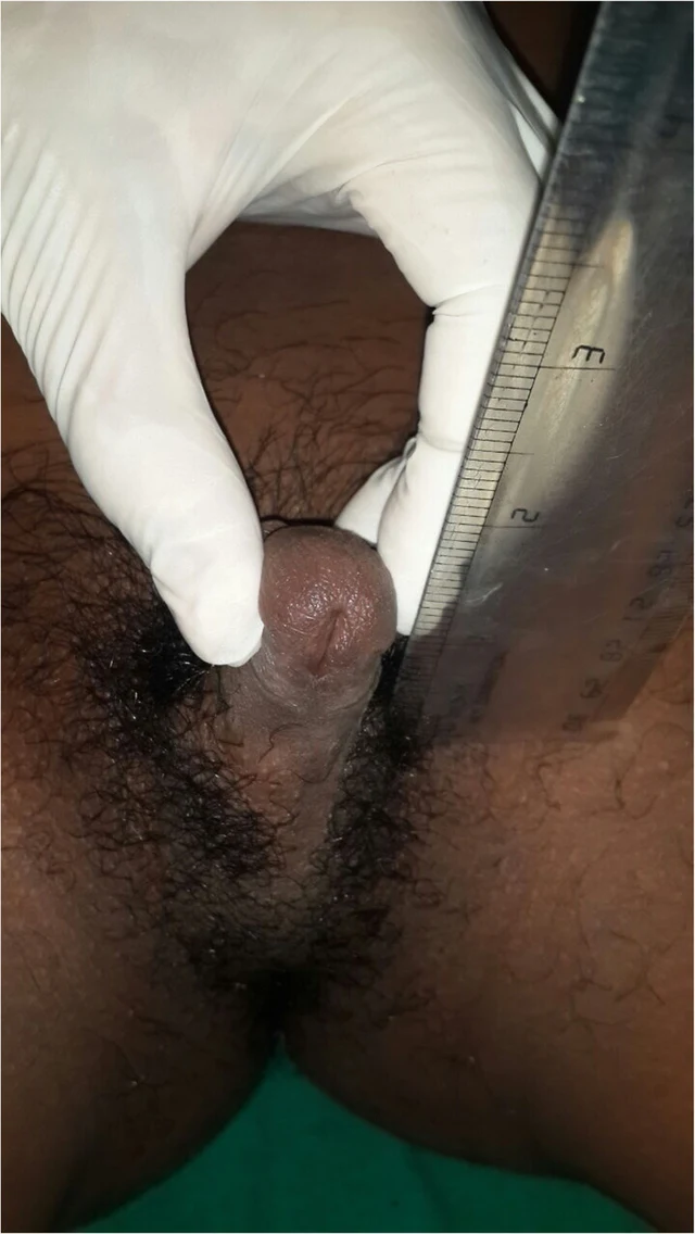
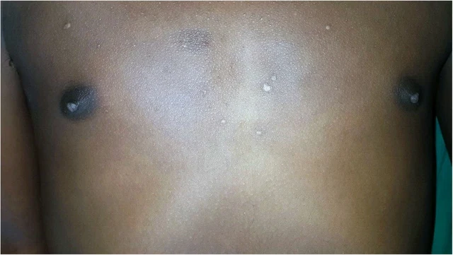
DSD-C01

Овотестикулярный DSD · 46,XX · DSD-C01

46,XX · HPO:0000023, HPO:0000047, HPO:0000062, Inguinal hernia (HP:0000023), Ambiguous genitalia (HP:0000062), Hypospadias (HP:0000047)

Правосторонний биполярный овотестис, ткань яичника и яичка примерно по 50%; при патологическом исследовании опухоли не выявлено, оценка фертильного потенциала тестикулярной ткани — 0.
СлучайПервоисточникАтлас
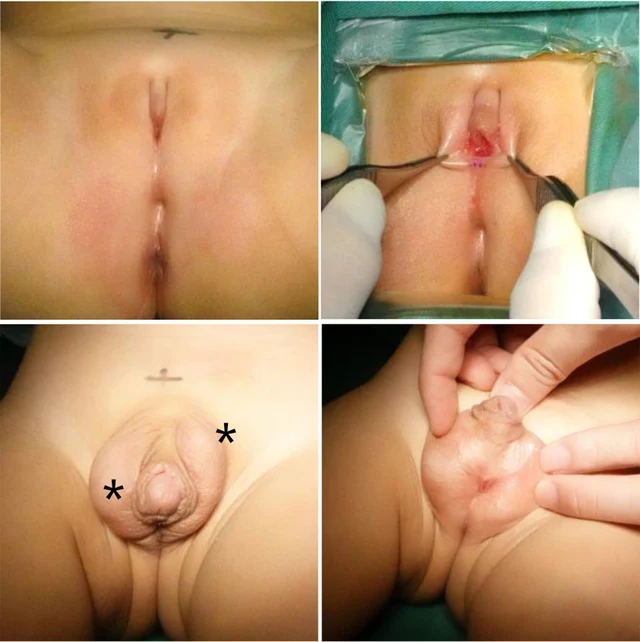
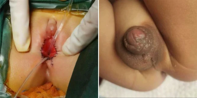
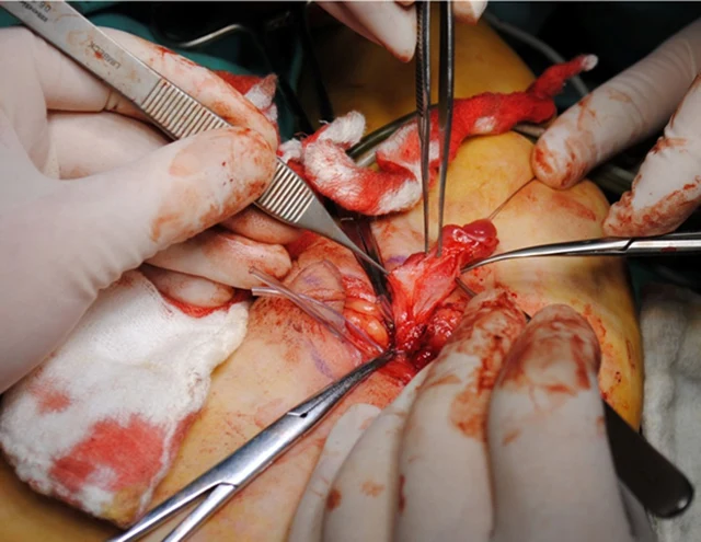
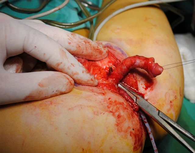
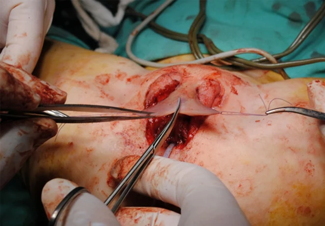
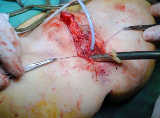
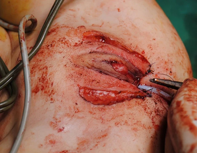
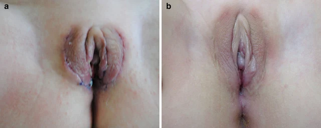
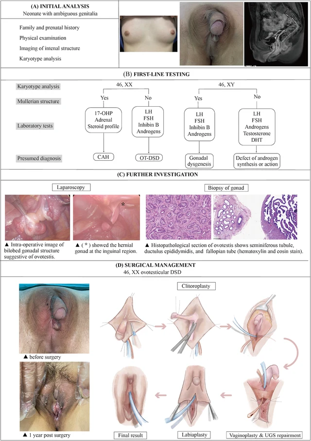
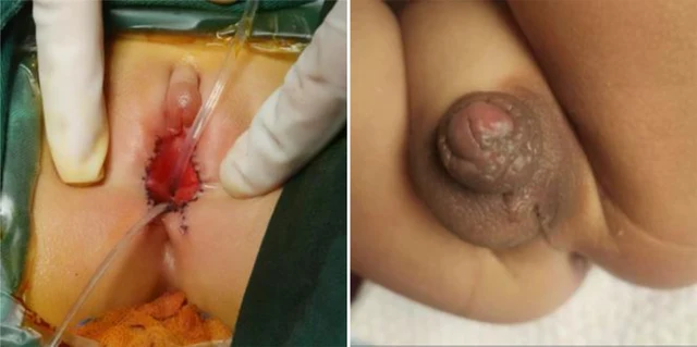
FIGURE 4 The illustration of the systematic diagnostic and treatment process for OT-DSD. The investigation process of OT-DSD can be summarized as initial assessment (A), first-line testing (B), and further investigation (C,D) showed the procedures of feminizing genitoplasty and pre- and 1-year post- operative pictures. The procedures included clitoroplasty, vaginoplasty and urogenital sinus repair, along with labiaplasty. 17, OHP, 17-alpha hydroxy progesterone; LH, Luteinizing hormone; FSH, follicle-stimulating hormone; DHT, dihydrotestosterone; CAH, congenital adrenal hyperplasia; OT-DSD, ovotesticular disorder of sex development. Zhao et al. 10.3389/fsurg.2024.1502340 Frontiers in Surgery 06 frontiersin.org 4 Изображения
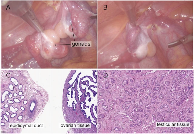
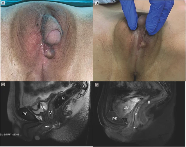
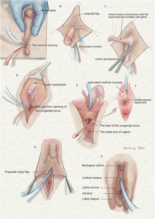
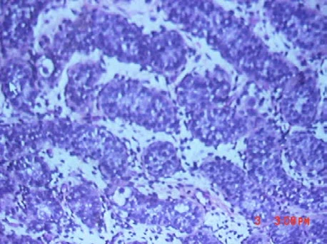
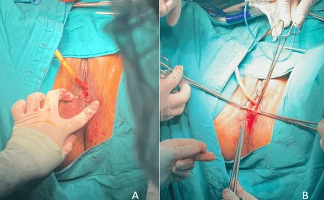
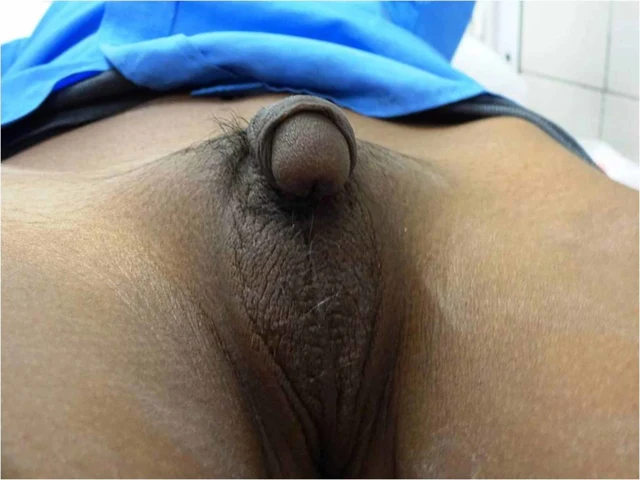
FIGURE 2 Intraoperative findings from laparoscopic exploration. (A) Intra-operative image of heterogenous gonadal structure showed the testicular and ovarian fractions organized in an end-to-end fashion, suggesting the ovotestis. (B) The right gonad was found at the inner ring of inguinal canal (*). (C) A biopsy was taken from the right hernial gonad in order to rule out any dysgenesis or ovotesticular tissue. Histopathological examination of sample 1 revealed epididymal and ovarian tissue. (D) Histopathological examination of sample 2 showed testicular histology with seminiferous tubules. Zhao et al. 10.3389/fsurg.2024.1502340 Frontiers in Surgery 03 frontiersin.org FIGURE 1 (A) Phenotypic appearance of genitalia before surgery. An enlarged clitoris, completely fused labia majora, and a movable painless mass at the right inguinal region was found. (B) An opening of the urogenital sinus was seen at the base of the clitoris. (C,D) Preoperative contrast-enhanced MRI showing well-defined urogenital sinus. Uterus was not detected. Sagittal MRI scan showing urinary bladder (UB), vagina (V), the common opening of the urogenital sinus (arrow), and rectum (R) The distance between the conference point to the superficial perineal pouch was below the level of the distal end of pubic symphysis (PS). Zhao et al. 10.3389/fsurg.2024.1502340 Frontiers in Surgery 02 frontiersin.org FIGURE 3 The illustration of the surgical procedures of feminizing genitoplasty. (A) The vulva showed Prader 4 virilization. (B) A subcoronal circumferential incision was made to de-glove the clitoris over Buck’s fascia. (C) The corporal bodies were excised with preservation of the glans clitoris and neurovascular bundles. (D) The corpora cavernosa was folded and sutured to the pubic arch. (E) The orange urethral catheter entered from the orifice through the urogenital sinus to exit from the fistula. (F) The fistula was sutured and secured with the rotating urethral mucosa flap. The vagina was pull-down to the vulve and sutured to the skin. (G) The prepuce in an onlay fashion covers the area between the clitoris and the vestibule. (H) The final result of the procedure. Zhao et al. 10.3389/fsurg.2024.1502340 Frontiers in Surgery 05 frontiersin.org

DSD-C011

46,XX овотестис-ДСП (OT-DSD). · 46,XX · DSD-C011

46,XX · HPO:0000047, Hypospadias (HP:0000047)

Дальнейшие диагностические исследования: уретроскопия подтвердила данные МРТ. Междисциплинарный консилиум: обсуждение в формате MDT с акцентом на диагностику и хирургическую тактику, с участием отделений пластической хирургии, эндокринологии, гинекологии, урологии, психологической и социальной службы.
СлучайПервоисточникАтлас
FIGURE 1 Genitalia abnormalities (a) & (b) External genitalia at 3 weeks. (c) Genitocystography. (d) Testicular biopsy a b c d 1 Изображения

DSD-C012

46,XY ДСП (дисгенезия гонад, интерстициальная делеция 8p23.1). · 46,XY · DSD-C012

AR, GATA4, SRD5A2, WT1 · 46,XY · HPO:0000028, HPO:0000047, HPO:0000054, Micropenis (HP:0000054), Hypospadias (HP:0000047), Cryptorchidism (HP:0000028)

Диагноз установлен только методом высокодифференцированного кариотипирования: интерстициальная делеция сегмента 8p23. Эта делеция часто встречается у пациентов с психомоторной задержкой и пороками сердца, но обычно не наблюдается у пациентов с первичными мочеполовыми аномалиями; по данным литературы, такие аномалии у мальчиков описаны почти в каждом втором случае.
СлучайПервоисточникАтлас
FIGURE 3: (A, B) CO2 Laser Vaginoplasty Psychological support was provided due to the potential impact of this condition on personal identity, perceptions of fertility, and sexual life. The psychological intervention helped the patient cope with the emotional and social challenges associated with the diagnosis, significantly improving her emotional well- being (Figure 4). 2024 Suárez Garcia et al. Cureus 16(11): e72895. DOI 10.7759/cureus.72895 5 of 9 1 Изображения

DSD-C013

Другой/неопределённый ДСП · 46,XX · DSD-C013

46,XX · HPO:0000135, Hypogonadism (HP:0000135)

При выполнении пробы Вальсальвы выявлено заращение девственной плевы, подтверждённое введением тампона-зонда и пальпацией. Пациентка направлена к генетику для уточнения дифференциального диагноза, включая мозаичную форму синдрома Тёрнера и заращение девственной плевы.
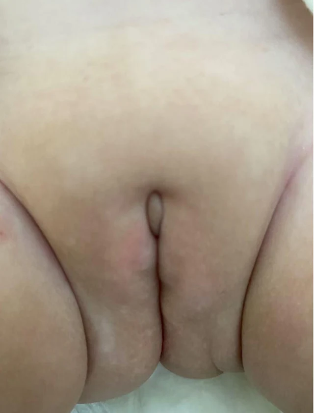
СлучайПервоисточникАтлас
Figure 1 The characteristics of the external genitalia of the proband at the age of 5 months. Ambiguous external genitalia was observed in the proband, with a 0.5 cm penis, a urethral opening in the perineum, a split scrotum resembling labia majora, and testicles found in the inguinal region. 1 Изображения

DSD-C014

46,XY ДСП · 46,XY · DSD-C014

NR0B1 · 46,XY

46,XY ДСП, связанный с дупликацией Xp21, в сочетании с мышечной дистрофией.
СлучайПервоисточникАтлас
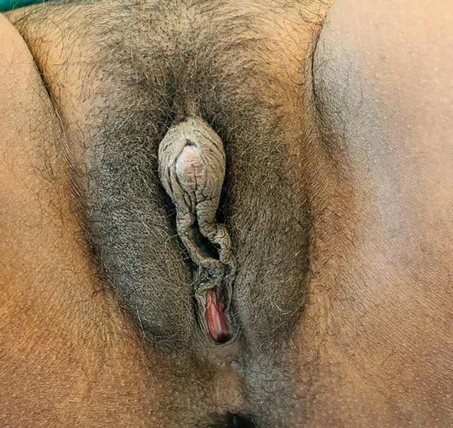
Figure 1 Clitoromegaly of 3 cm length and 1 cm breadth with clitoral index of 300 m2. 2 JCEM Case Reports, 2026, Volume 4, Issue 7 1 Изображения

DSD-C015

Врождённая гиперплазия коры надпочечников · 46,XX · DSD-C015

CYP21A2 · 46,XX · HPO:0004322, Short stature (HP:0004322)

После первичного диагноза родители больше не обращались за повторной консультацией по поводу аменореи. Диагностическая оценка: при осмотре низкий рост (140,5 cm; медиана роста родителей 151 cm, −3,1 SDS), масса тела 42 kg, индекс массы тела 21 kg/m², артериальное давление и пропорции тела в норме, стадии Tanner A4B3P4.
СлучайПервоисточникАтлас
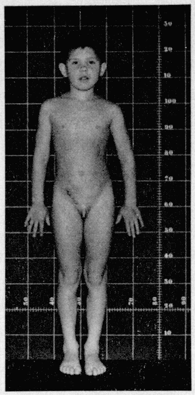
Patient with POR deficiency at 9 years old and her external genitalia appearance. 5 Изображения
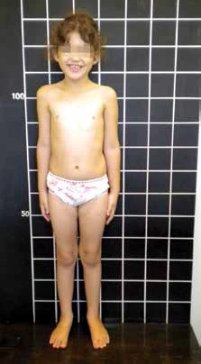
Full-body photograph of the 9-year-old girl with POR deficiency (stature grid background). External genitalia appearance of the POR deficiency patient. "Hammered silver" appearance associated with craniosynostosis (face at 9 years). Figure 1. Mother´s (A) and child’s (B) external genitalia 8 months after delivery. A B

DSD-C016

46,XX DSD; дефицит цитохром P450-оксидоредуктазы (POR) в сочетании с синдромом Антли-Бикслера. · 46,XX · DSD-C016

46,XX · HPO:0000135, Hypogonadism (HP:0000135)

Таким образом, исходя из тяжёлой вирилизации матери во время беременности, в качестве основной диагностической гипотезы рассматривалась недостаточность CYP19. Однако результаты анализа гена CYP19 этот диагноз не подтвердили.
СлучайПервоисточникАтлас
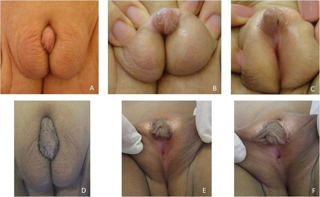
Fig. 2 The external genitalia of our patient at 10 months (A, B, C) and at 6 years old (D, E, F) 1 Изображения

DSD-C017

46,XY DSD (дефицит 17β-гидроксистероиддегидрогеназы 3-го типа, дефицит HSD17B3). · 46,XY · DSD-C017

AR, HSD17B3, NR5A1, SRD5A2 · 46,XY

46,XY DSD, обусловленный дефицитом HSD17B3.
СлучайПервоисточникАтлас
FIGURE 1: Clinical photograph showing fused labioscrotal folds with absent gonads, ventral chordee, and penoscrotal hypospadias Anthropometry revealed a weight of 17 kg, a height of 101 cm (more than 75th centile) with midparental height of 169 cm for a male child (between 25th and 50th centile) and 156 cm for a female child (less than 5th centile). Investigations revealed hypokalemia, primary adrenal insufficiency with elevated sex steroids, and elevated 11-deoxycortisol ( 2 Изображения
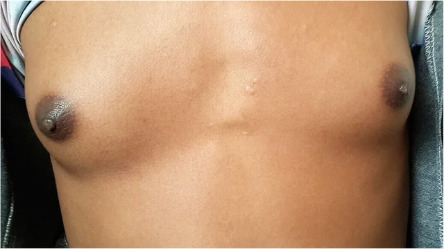
FIGURE 2: Clinical photograph showing Tanner stage 3 breast development There was no complaint of cyclical abdominal pain or hematuria. At this stage, the child was re-admitted and underwent definitive hysterectomy and bilateral salpingo-oophorectomy. This was followed by subcutaneous mastectomy and chordee correction. The child is now planned for a second stage surgery for hypospadias correction, following which testosterone replacement therapy will be initiated.

DSD-C018

46,XX DSD (врождённая гиперплазия коры надпочечников, дефицит 11β-гидроксилазы). · 46,XX · DSD-C018

46,XX · HPO:0000047, Hypospadias (HP:0000047)

Ребёнка воспитывали в семье как мальчика и дали ему соответствующее мужское имя. При физикальном осмотре артериальное давление в положении лежа составило 144–154/90–94 mmHg (выше 99-го процентиля для данного возраста и роста, что соответствует диагнозу артериальной гипертензии).
СлучайПервоисточникАтлас
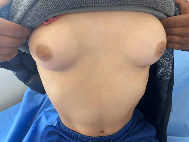
FIGURE 1: Clinical image showing breast development during central precocious puberty (Tanner stage 4). 2025 Hamaichat et al. Cureus 17(5): e83965. DOI 10.7759/cureus.83965 3 of 7 2 Изображения
FIGURE 2: Clinical image showing ambiguous genitalia at Prader stage 5 and pubic hair at Tanner stage 5. Parameter Value Weight -1.5 SD Height -2 SD Pubic hair Tanner stage 5 Breast Tanner stage 4 External genitalia Prader stage 5 Follicle-stimulating hormone 4.48 mIU/mL (normal range: 2–13 mIU/mL) Luteinizing hormone 2.04 mIU/mL (normal range: 1.5–8 mIU/mL) Estradiol 52.23 pg/mL (34–186 pg/mL) Pelvic ultrasound Pubertal-sized uterus and a left lateral uterine structure 11 mm in height (left ovary), with the right ovary not visualized

DSD-C019

46,XX DSD (врождённая гиперплазия коры надпочечников, дефицит 11β-гидроксилазы). · 46,XX · DSD-C019

CYP11B1 · 46,XX · HPO:0000047, Hypospadias (HP:0000047)

Врождённая гиперплазия коры надпочечников (дефицит 11β-гидроксилазы); 46,XX женщина, вирилизация наружных гениталий Prader 5 степени, преждевременное половое развитие по гетеросексуальному типу.
СлучайПервоисточникАтлас
Клиническое изображение · DSD-C02 3 Изображения
Клиническое изображение · DSD-C02 Клиническое изображение · DSD-C02

DSD-C02

Врожденная гиперплазия коры надпочечников · 46,XX · DSD-C02

46,XX · HPO:0000047, HPO:0000771, Hypospadias (HP:0000047), Gynecomastia (HP:0000771)

Авторы на основании артериальной гипертензии, вирилизации и результатов исследования гипоталамо-гипофизарно-надпочечниковой оси клинически диагностировали дефицит 11β-гидроксилазы; генетическое подтверждение и определение 11-дезоксикортизола не проводились.
СлучайПервоисточникАтлас
Figure 5 Postoperative appearance of the external genitalia of twins. (A) Twin A at 30 months of age after feminizing surgery. (B) Twin B at 31 months of age after stage II hypospadias repair. associated with DSD, a group of rare conditions in which the chromosomal, gonadal, or phenotypical sex is incongruent (8). We herein present a case of 46,XY identical twins with DSD and UBMS caused by a novel heterozygous de novo splicing variant (c.1551-2A>G) in PPP1R12A. This variant results in the presence of a uterus in both 46,XY twins. Twin A exhibited gonadal dysgenesis and was assigned female, while Twin B had functional testes and was raised as male. PPP1R12A encodes PPP1R12A (1030aa), also named myosin phosphatase-targeting subunit 1 (MYPT1), expressed extensively and strongly in the brain, thyroid, 2 Изображения
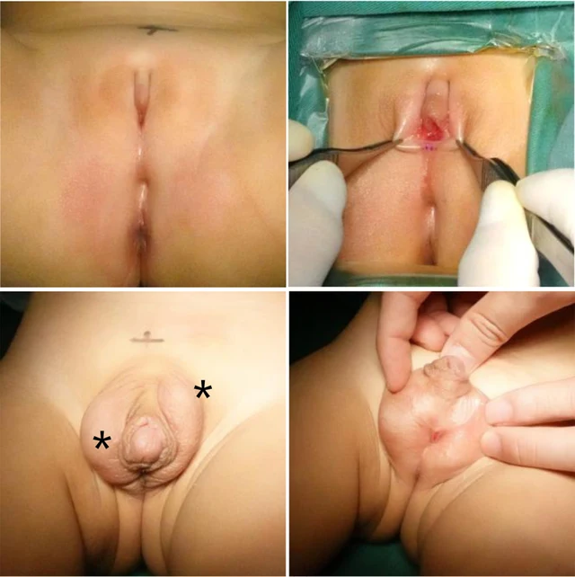
Figure 1 Appearance of the external genitalia of twins. (A,B) Twin A at 10 months of age. (C,D) Twin B at 30 months of age after stage I hypospadias repair. Testis-like masses were palpable in the area of stars.

DSD-C020

46,XY DSD · 46,XY · DSD-C020

46,XY

46,XY DSD, связанный с вариантом PPP1R12A, с аномалиями мочеполового тракта и гонадной дисгенезией.
СлучайПервоисточникАтлас
Figure 1 Appearance of the external genitalia of twins. (A,B) Twin A at 10 months of age. (C,D) Twin B at 30 months of age after stage I hypospadias repair. Testis-like masses were palpable in the area of stars. 2 Изображения
Figure 5 Postoperative appearance of the external genitalia of twins. (A) Twin A at 30 months of age after feminizing surgery. (B) Twin B at 31 months of age after stage II hypospadias repair. associated with DSD, a group of rare conditions in which the chromosomal, gonadal, or phenotypical sex is incongruent (8). We herein present a case of 46,XY identical twins with DSD and UBMS caused by a novel heterozygous de novo splicing variant (c.1551-2A>G) in PPP1R12A. This variant results in the presence of a uterus in both 46,XY twins. Twin A exhibited gonadal dysgenesis and was assigned female, while Twin B had functional testes and was raised as male. PPP1R12A encodes PPP1R12A (1030aa), also named myosin phosphatase-targeting subunit 1 (MYPT1), expressed extensively and strongly in the brain, thyroid,

DSD-C021

46,XY DSD (различия полового развития) · 46,XY · DSD-C021

46,XY · HPO:0000047, Hypospadias (HP:0000047)

46,XY DSD, связанный с вариантом PPP1R12A, с аномалиями мочеполового тракта.
СлучайПервоисточникАтлас
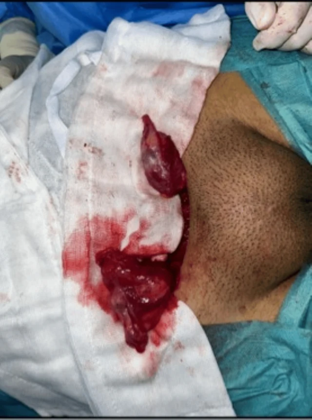
FIGURE 4: Intraoperative appearance of bilateral gonadal structures. Surgical exploration demonstrating dysgenetic gonadal tissue located in the inguinal region. Histopathological examination revealed dysgenetic testicular tissue with a limited number of medium-sized seminiferous tubules showing maturation arrest at the primary spermatocyte stage, sparse Leydig cells, and absence of ovarian parenchyma or stroma. These findings were consistent with dysgenetic testicular tissue. Management and follow-up Management was discussed in a multidisciplinary setting involving pediatric endocrinologists, pediatric surgeons, geneticists, and psychologists. Age-appropriate information was provided, and shared decision- making was conducted with the patient and her parents. The patient expressed a stable female gender identity. Bilateral orchiectomy was performed in accordance with the declared gender, with psychological support. Histological analysis of the resected gonads showed complete testicular atrophy without intratubular germ cell neoplasia or malignant features. Estrogen replacement therapy with 17β-estradiol was subsequently initiated, along with management of hyperandrogenism and regular endocrinological follow-up. 1 Изображения

DSD-C022

46,XY DSD (различия полового развития) · 46,XY · DSD-C022

NR5A1 · 46,XY · HPO:0000135, Hypogonadism (HP:0000135)

Параметры роста: рост 164 cm (+1.5 SD), масса тела 75 kg (>+2 SD), индекс массы тела 28.2 kg/m², что согласно референсным кривым роста для девочек соответствует избыточной массе тела.
СлучайПервоисточникАтлас
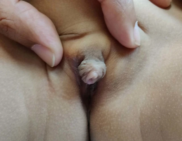
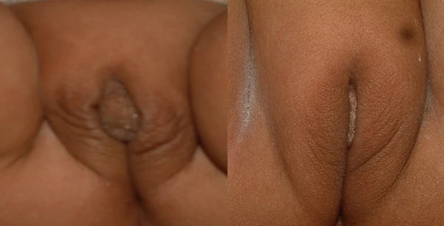
Figure 1 Genital appearance at 4 months showing coarse rugosity and visibly enlarged clitoris, and then at 2 years of age showing fine rugosity over labia and clitoral enlargement partially covered by labial fullness (patient K). 1 Изображения

DSD-C023

Врождённая гиперплазия коры надпочечников · DSD-C023

Несмотря на выявленную при рождении неопределённость наружных гениталий, диагноз CAH (врождённой гиперплазии коры надпочечников) был установлен, а терапия стероидами начата только в 9 месяцев. У её двух сестёр 15 и 5 лет и одного брата 11 лет также диагностирована классическая вирильная форма CAH.

СлучайПервоисточникАтлас
Figure 2 Genital appearance at age 3.2 years and then at 4 years showing almost normal female genitalia (patient N). Congenital adrenal hyperplasia 557 1 Изображения

DSD-C024

Врождённая гиперплазия коры надпочечников · DSD-C024

Несмотря на выявленную при рождении неопределённость наружных гениталий, диагноз CAH (врождённой гиперплазии коры надпочечников) был установлен, а терапия стероидами начата только в 9 месяцев. У её двух сестёр 15 и 5 лет и одного брата 11 лет также диагностирована классическая вирильная форма CAH.
СлучайПервоисточникАтлас
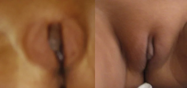
Figure 3 Genital appearance at initial presentation (2.5 years) and 1 year later (patient A). 558 B. Kulshreshtha et al. 1 Изображения

DSD-C025

Врождённая гиперплазия коры надпочечников · DSD-C025

Несмотря на выявленную при рождении неопределённость наружных гениталий, диагноз CAH (врождённой гиперплазии коры надпочечников) был установлен, а терапия стероидами начата только в 9 месяцев. У её двух сестёр 15 и 5 лет и одного брата 11 лет также диагностирована классическая вирильная форма CAH.
СлучайПервоисточникАтлас
Figure 4. Appearance of external genitalia of the patient with features of partial virilization, including a small phallic structure, partially fused labioscrotal folds, and an external urethral meatus located in the perineal region, below the opening of the surgically created vagina. 1 Изображения

DSD-C026

46,XY DSD (частичная нечувствительность к андрогенам в сочетании с врождённой гиперплазией коры надпочечников, дефицит P450-оксидоредуктазы). · 46,XY · DSD-C026

46,XY

Представлено клиническое наблюдение пациентки 26 лет, у которой в раннем грудном возрасте в связи с неоднозначным строением мочеполовых органов был диагностирован DSD. Данные опроса о происхождении ребёнка не выявили патологий, значимых для диагностики.
СлучайПервоисточникАтлас
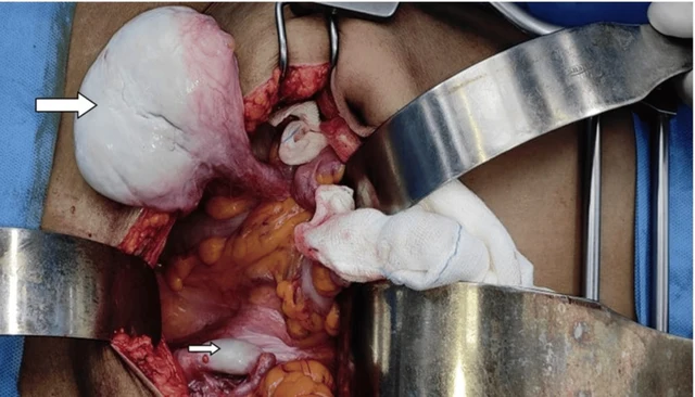
FIGURE 4: Testicular mass being separated from the local structures. Big arrow: Right testis, Small arrow: Left testis 2026 Ramya Lakshmi et al. Cureus 18(4): e106608. DOI 10.7759/cureus.106608 4 of 9 3 Изображения
FIGURE 1: Absence of pubic hair growth FIGURE 2: Absence of axillary hair 2026 Ramya Lakshmi et al. Cureus 18(4): e106608. DOI 10.7759/cureus.106608 2 of 9

DSD-C027

Синдром нечувствительности к андрогенам · 46,XY · DSD-C027

AR · 46,XY · HPO:0000786, Primary amenorrhea (HP:0000786)

Полная нечувствительность к андрогенам (CAIS); злокачественная герминогенная опухоль из внутрибрюшного яичка (дисгерминома/семинома, TxN3M0S1, стадия IIC); первичная аменорея; отсутствие матки.
СлучайПервоисточникАтлас
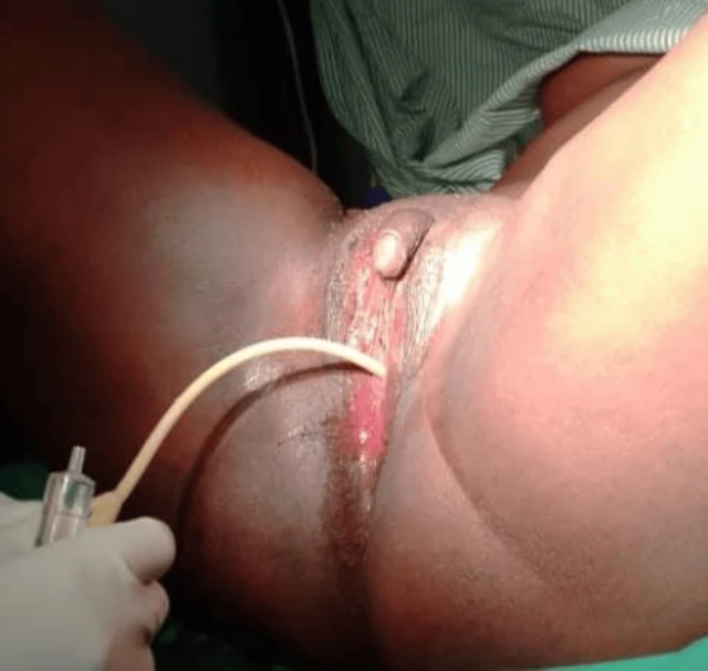
FIGURE 2: Case one catheterized At the six-week follow-up, the patient reported marked improvement in psychological well-being and satisfaction with the surgical outcome. Examination demonstrated a well-healed perineum and a functional vaginal canal measuring approximately 7 cm in length and 3 cm in width, as shown in Figure 3. She was also reviewed at three months and six months postoperatively, and she was happily sexually active without a complaint. Physical examination showed a long wide vaginal opening of about 7 cm long by 3 cm wide. 2026 Ainomugisha et al. Cureus 18(6): e111019. DOI 10.7759/cureus.111019 4 of 11 3 Изображения
FIGURE 3: Case one six weeks post-surgery Case two A 25-year-old individual raised and socially recognized as male presented with progressive abdominal distension and lower abdominal pain of one year duration. During clinical evaluation, the patient disclosed the onset of virilization during adolescence, including the development of facial hair and a deepened voice, but also reported regular menstruation beginning at 15 years of age. The patient had been assigned male gender at birth because of the presence of a phallus, which had enlarged progressively with age. However, urine did not pass through the phallus. The patient identified socially as male and expressed a desire for relief from the abdominal swelling and cessation of menstruation. Both parents were deceased, and the patient was accompanied by a community health worker in whom he had previously confided. He had no formal education and survived on casual labor. On examination, the patient appeared chronically ill and wasted but demonstrated masculine body habitus, facial hair, a hoarse voice, smooth skin, and well-developed breasts. Vital signs were stable. Abdominal examination revealed gross distension with marked suprapubic tenderness. A large solid pelvic mass measuring approximately 18 × 10 cm was palpable, in addition to another irregular mass within the left lumbar region. Bilateral inguinal nodular swellings were also noted. Perineal examination demonstrated a phallus measuring approximately 6 × 3 cm without a urethral opening. A closed introitus with a small distal dimple was observed, through which urine was passed with straining in the lithotomy position. The stage, according to the Prader score, was 2. The anal opening was normally positioned. A provisional diagnosis of 2026 Ainomugisha et al. FIGURE 1: Case one before surgery A provisional diagnosis of ambiguous genitalia with suspected 46, XX DSD was made. Pelvic ultrasonography demonstrated a normal uterus, cervix, and right ovary. A cystic lesion measuring 33 × 4 × 20 mm with multiple internal echoes and a thick wall was identified within the left ovary. No abnormalities were detected in the urinary tract or upper abdominal organs. Due to financial limitations, karyotyping and hormonal assays could not be performed. The patient expressed a strong desire for surgical correction of the vaginal obstruction and removal of the enlarged clitoris after extensive counseling regarding anatomy, sexual function, and potential risks of surgery. Surgical Procedure Under spinal anesthesia and lithotomy positioning, examination under anesthesia confirmed a normal urethral opening located superior to the vaginal dimple. Catheterization, as shown in Figure 2, verified that the enlarged clitoris was not associated with the urinary tract. A diamond-shaped incision was created at the vaginal opening to enlarge the introitus. A vaginal canal approximately 6 cm long and 2 cm wide was identified, and the cervix was visualized. Vaginal mucosa was mobilized and anchored to the labial and perineal skin edges. A 20-mL syringe barrel was inserted and secured as a temporary vaginal stent. Clitoral reduction was subsequently performed by excision at the base near the mons pubis, followed by reconstruction and layered closure with meticulous hemostasis. Postoperatively, the patient remained hospitalized for 10 days and received antibiotics, analgesics, and perineal hygiene care. The urinary catheter 2026 Ainomugisha et al. Cureus 18(6): e111019. DOI 10.7759/cureus.111019 3 of 11

DSD-C028

46,XX DSD (нарушение развития пола) · 46,XX · DSD-C028

46,XX

Авторы сообщают о неоднозначных гениталиях, подозрение на 46,XX DSD.
СлучайПервоисточникАтлас
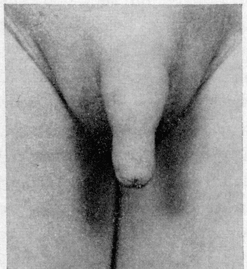
Fig. 1. 7 years old girl with female pseudohermaphroditism due to virilizing adenoma of the mother. 2 Изображения
Fig. 2. Male external genitalia of the same girl. The karyotype was 46XX. Serum electrolytes were within normal limits. The results of the steroid studies are presented below in more detail. A voiding cysto-urethrogram showed no anomalies. At surgery, the right inguinal canal was opened and no gonad was found. On revision of the abdominal cavity, two bean-sized gonads, two Fallopian tubes, and a small uterus attached to the bladder were detected. No Wolffian structures were found. A biopsy was taken from each gonad. Histological examination revealed ovarian tissue in both biopsies and 42 per cent of the

DSD-C029

46,XX DSD (женский ложный гермафродитизм, внутриутробное воздействие андрогенов вследствие аденомы коры надпочечников у матери). · 46,XX · DSD-C029

46,XX · HPO:0000028, Cryptorchidism (HP:0000028)

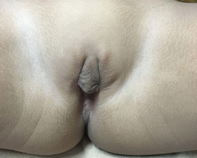
У пациентки диагностирован женский ложный гермафродитизм (46,XX, полная маскулинизация), обусловленный секрецией андрогенов доброкачественной аденомой коры правого надпочечника матери; морфологический диагноз у матери — доброкачественная аденома коры надпочечников (65 г, 6,5×5×3 см, злокачественных клеток нет).
СлучайПервоисточникАтлас
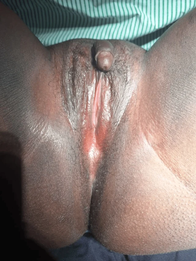
Клиническое изображение · DSD-C03 2 Изображения
Клиническое изображение · DSD-C03

DSD-C03

Дефицит ароматазы · 46,XX · DSD-C03

46,XX

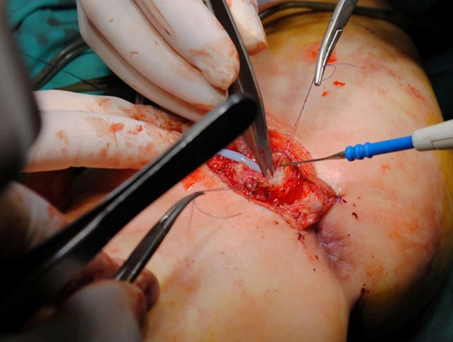
С учетом указаний на вирилизацию во время беременности у матери, кариотипа 46,XX и исключения распространенных надпочечниковых/тестикулярных источников авторы считают дефицит ароматазы вероятным.
СлучайПервоисточникАтлас
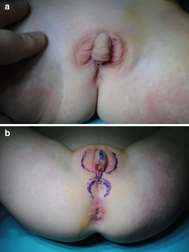
Fig. 4 Surgical management of ambiguous genitalia: third step, creation of a vagina 8 Изображения
Fig. 1 a The preoperative aspect of ambiguous genitalia. b The design of further surgical incisions Fig. 3 Surgical management of ambiguous genitalia: second step, removing the erectile tissue from the clitoris Fig. 2 Surgical management of ambiguous genitalia: first step, clitoris degloving 550 Aesth Plast Surg (2014) 38:549–553 123 Fig. 6 Surgical management of ambiguous genitalia: fifth step, creation of a neovulva through tailoring of the labia minora using the dorsal clitoridean skin Aesth Plast Surg (2014) 38:549–553 551 123 Fig. 5 Surgical management of ambiguous genitalia: fourth step, retrourethral placement of the vagina Fig. 8 The aspect of ambiguous genitalia, postoperatively (a) and 2 years after surgery (b) 552 Aesth Plast Surg (2014) 38:549–553 123 Fig. 7 Surgical management of ambiguous genitalia: last step, fixing the flaps with separate sutures

DSD-C030

46,XX DSD (врождённая гиперплазия коры надпочечников, дефицит 21-гидроксилазы, классическая/сольтеряющая форма). · 46,XX · DSD-C030

46,XX

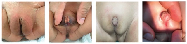
Анамнез и лечение: девочка 3 лет обратилась по поводу классической CAH (обусловленной дефицитом 21-гидроксилазы), диагностированной в 6 недель. Повышение сывороточного 17-OHP (150 нмоль/л) подтверждает сольтеряющую форму CAH.
СлучайПервоисточникАтлас
Figure 1: Patient’s abnormal genital phenotype: at the age of three days, (a) labia-majora-like bifid scrotum with clitoris-like microphallus and (b) perineoscrotal hypospadias; at the age of two years and three months, (c, d) microphallus had grown to 1.5 cm in length. 1 Изображения

DSD-C031

Дефицит 5α-редуктазы · 46,XY · DSD-C031

SRD5A2, SRY · 46,XY · HPO:0000047, HPO:0000054, Micropenis (HP:0000054), Hypospadias (HP:0000047)

46,XY DSD вследствие дефицита 5α-редуктазы-2; молекулярное исследование выявило комплексно-гетерозиготные варианты SRD5A2 c.485A>C (p.His162Pro) и c.680G>A (p.Arg227Gln).

СлучайПервоисточникАтлас
Загрузить ещё случаи

Листайте влево или вправо, чтобы просмотреть изображения этого случая

Открыть исходное изображение
СлучайПервоисточникАтласИсходная подпись на китайском
DSD Research Library Версия данных v1.4.0