 2 Images
46,XY DSD · 46,XY · DSD-C032
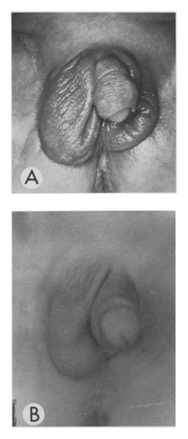
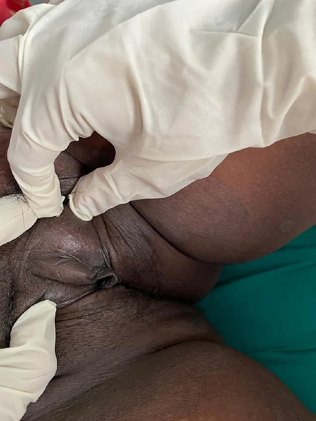
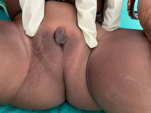
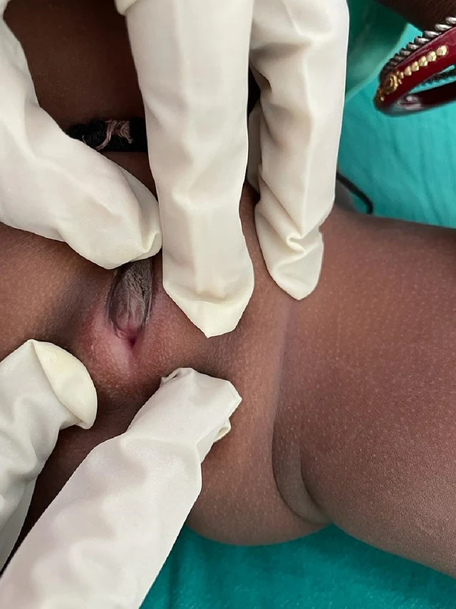
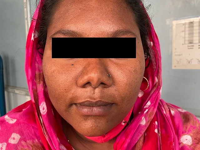
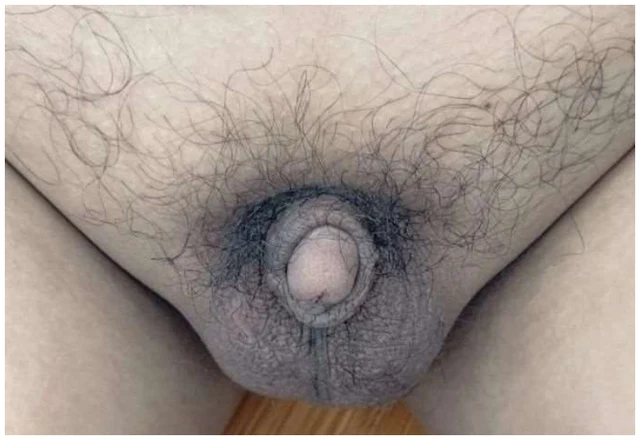
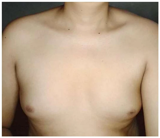
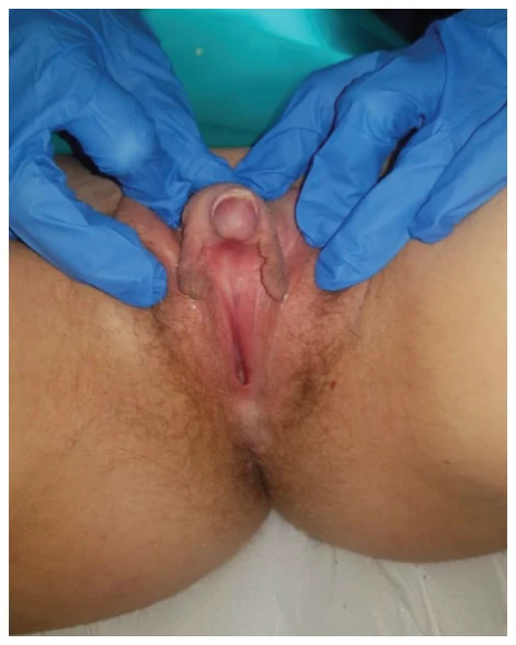
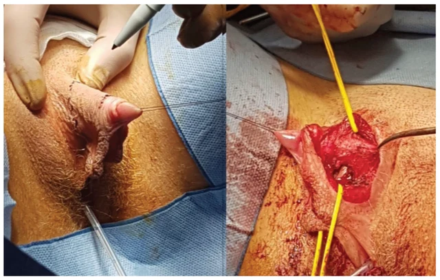
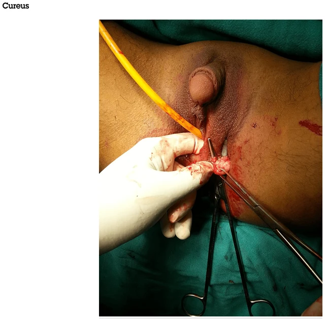
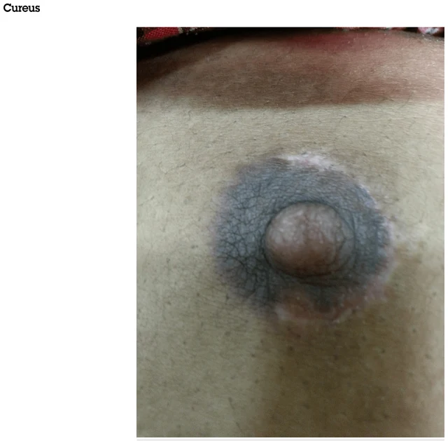
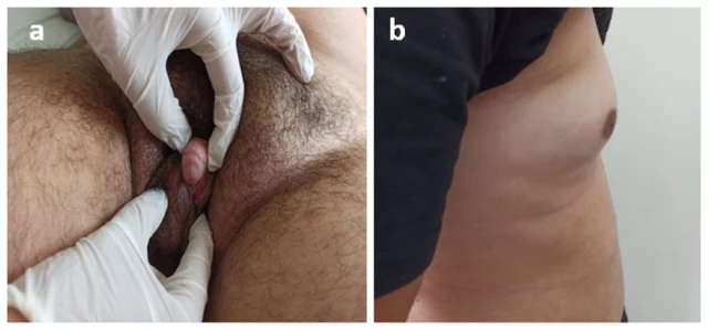
17β-HSD3 deficiency supported by low testosterone/androstenedione ratio and compound-heterozygous HSD17B3 variants.
Browse cases and figures from the original collection. Search by condition, gene, karyotype or phenotype and follow the source publication.
280 Image cases571 Images
Current selection: 280 Image cases · 571 Images
Select an image to enlarge it. Original images retain the complete source file.
2 Images
17β-HSD3 deficiency supported by low testosterone/androstenedione ratio and compound-heterozygous HSD17B3 variants.
 2 Images
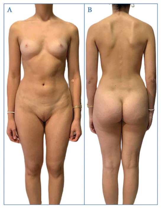
Complete androgen insensitivity syndrome (CAIS), supported by female phenotype, 46,XY karyotype, male-range androgen levels, absent uterus and intra-abdominal testes; histopathology showed atrophic undescended testes.
 1 Images
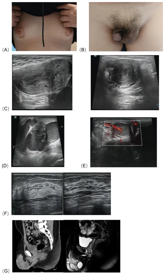
SRY-negative 46,XX testicular disorder of sex development with ectopic Müllerian structures; right gonadal biopsy confirmed testicular tissue.
 3 Images
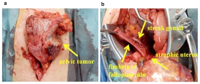
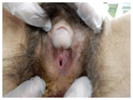
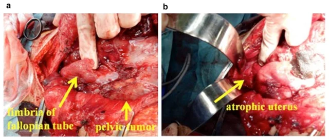
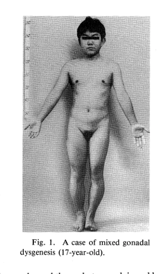
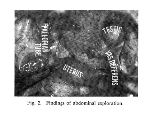
The final pathological diagnosis was “seminoma” (Fig. The karyotype was found to exhibit 45,X/46,XY mosaicism, and the patient was diagnosed with MGD.
 1 Images
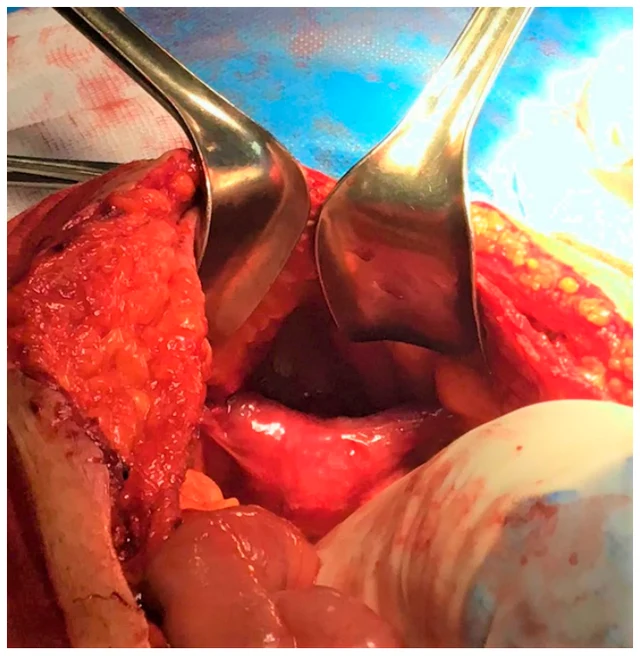
Based on the intraoperative findings, history of bilateral cryptorchidism, MRI imaging and karyotype, diagnosis of PMDS was made. It is very likely that this figure underestimates the true incidence of the syndrome as the diagnosis of PMDS largely depends on the intraoperative detection of Müllerian derivatives.
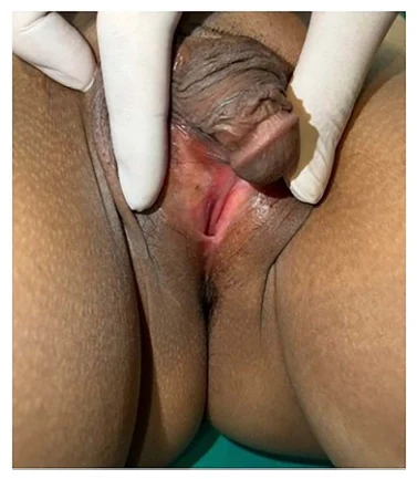 2 Images
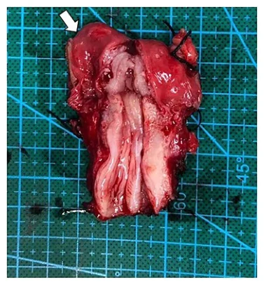
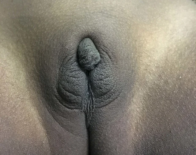
She was diagnosed as phenotypic female and reared as a girl without further evaluation.
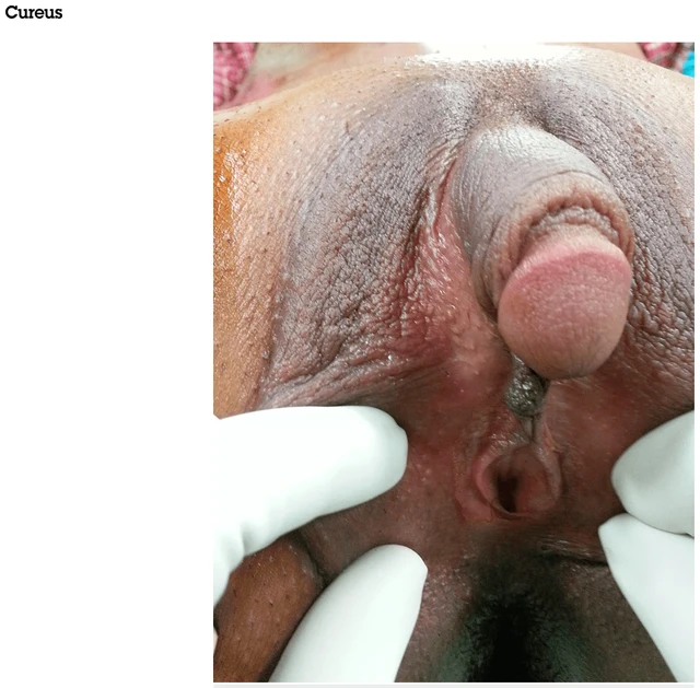 4 Images
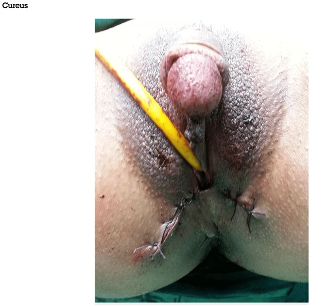
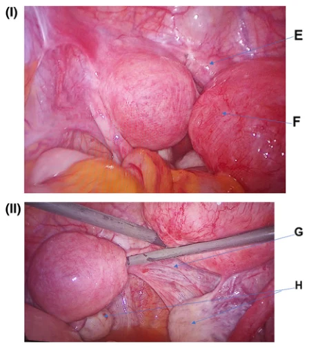
Both reports confirmed a normal uterus with bilateral normal-sized ovaries. Solid masses removed as bilateral testes were histologically confirmed as Bartholin gland hyperplasia (Figure 6).
 1 Images
There was a blind transverse connection between the two uteruses and a horseshoe band cervix, which confirmed the diagnosis of mullerian agenesis or Mayer–Rokitansky–Kuster– Hauser (MRKH) syndrome (Fig. Hence, it was confirmed to be type 1 MRKH syndrome.
 2 Images
Integrating the phenotype and the endocrine exclusion process, the authors clinically considered aromatase deficiency.
 1 Images
Sanger sequencing confirmed maternal inheritance in the proband, her healthy mother, and grandfather, consistent with CDKN1C paternal imprinting.
 1 Images
The patient was ultimately diagnosed with type II Leydig cell hypoplasia.
 2 Images
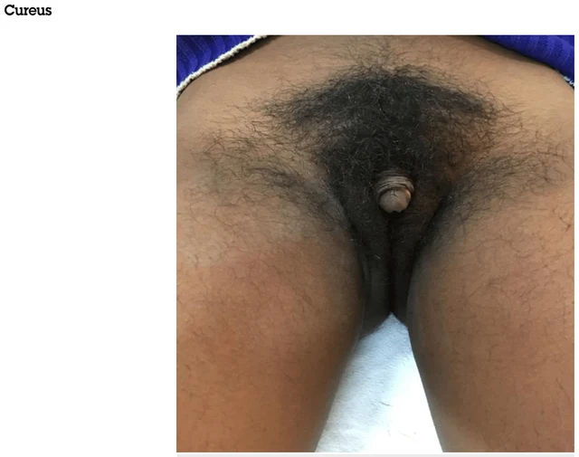
The diagnosis of CAH, the simple-virilizing type, was made. µg/dL; >700 µg/dL suggests androgen-secreting adrenal tumors) was raised but 17-hydroxyprogesterone (17-OHP) at 55 ng/dL (<200 ng/dL excludes the diagnosis; >800 ng/dL is diagnostic, intermediate result require adrenocorticotropic hormone (ACTH) stimulation test) was low, which was against the diagnosis of CAH.
 1 Images
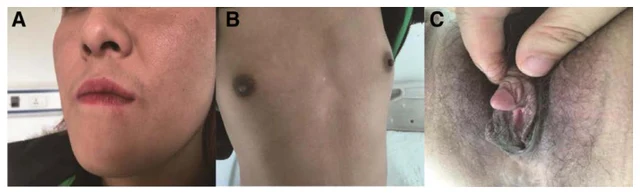
She was diagnosed with GD 2 months before admission to our hospital due to palpitation, heat intolerance, muscle weakness. Follow-up We confirmed the coexistence of SV 21-OHD, GD and 47, XXX.
 1 Images
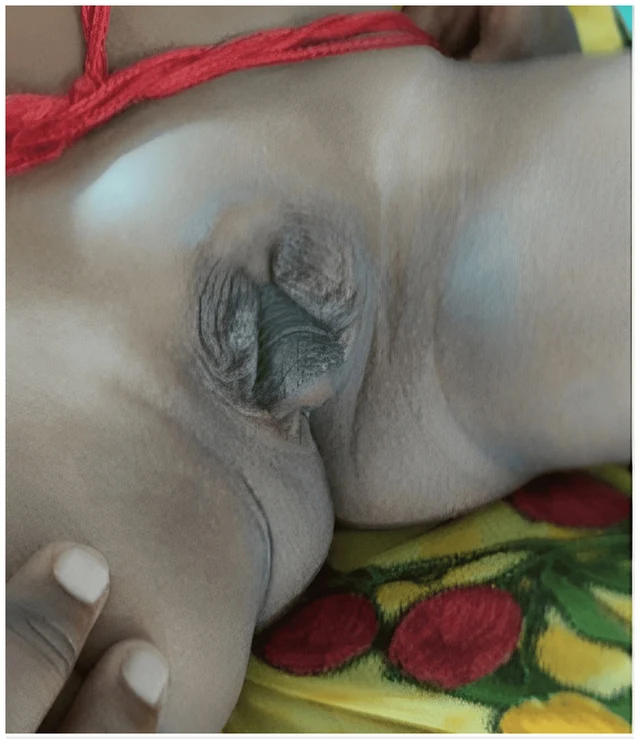
de la Chapelle syndrome / 46,XX testicular DSD
 1 Images
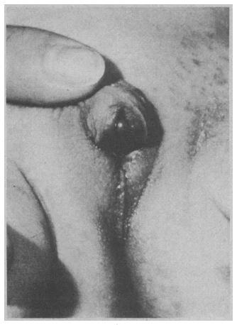
Silver-Russell syndrome with external genitalia anomalies
 2 Images
 1 Images
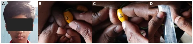
Normosmic hypogonadotropic hypogonadism with short stature
 2 Images
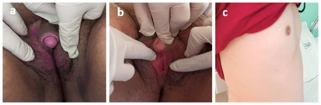
Classic simple virilizing CAH due to 21-hydroxylase deficiency
 1 Images
Swyer syndrome / 46,XY complete gonadal dysgenesis
 1 Images
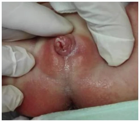
X-linked lissencephaly with ambiguous genitalia (X-LAG)
 1 Images
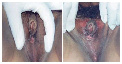
P450 oxidoreductase deficiency (PORD)